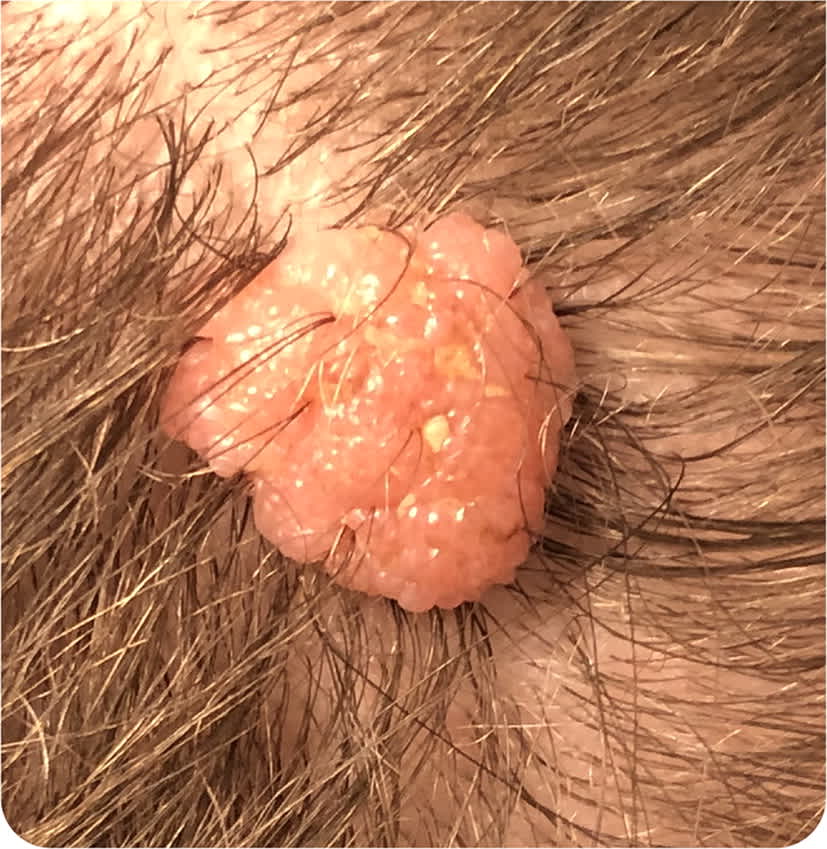
A 17-year-old patient presented with a lump on the head. The patient first noticed the lump at 11 years old. It had been slowly growing over the previous few years. The lump was not painful and did not bleed. It occasionally itched and bothered the patient when they brushed their hair. The patient did not recall injury or trauma to the area. Physical examination revealed a skin-colored polypoid mass on the top of the patient's head, above the parietal area (Figure 1). The mass was raised and asymmetrical with slightly irregular borders. It measured 14 mm × 12 mm in size.
FIGURE 1
Question
Based on the patient's history and physical examination findings, which one of the following is the most likely diagnosis?
- A. Acrochordon.
- B. Compound nevus.
- C. Dermal nevus.
- D. Junctional nevus.
Discussion
The answer is C: dermal nevus. Commonly called moles, nevi are benign tumors composed of nevus cells. They can be acquired or congenital. Nevi appear on 1% to 2% of newborns and increase in incidence throughout life, peaking in the 30s and 40s. Most nevi are benign. However, some, such as large congenital nevi and atypical moles, may become malignant. Congenital nevi are considered large in adults if they are at least 20 cm in diameter and in neonates if they are at least 9 cm on the head or neck or 6 cm on other areas.1–4
The three subtypes of nevi (compound, dermal, and junctional) are differentiated by the location of the nevus cells in the skin. Nevus cells typically start in the dermoepidermal junction and migrate into the dermis. During the transition, a lesion is considered a compound nevus. The development is sequential, and progression can stop at any point.3,5
The shape and size of dermal nevi vary widely. They can be warty, polypoid, or pedunculated. Like the other subtypes, dermal nevi can be hyperpigmented or pink or appear the same color as surrounding skin.1,5
Acrochordons are commonly known as skin tags. They are usually the color of surrounding skin but can be hyperpigmented. They are often pedunculated and range from 2 mm to 5 mm in size but can be larger. They increase in number with age and during pregnancy.6
Compound nevi are usually hyperpigmented or the color of surrounding skin with a smooth and elevated or warty surface. Elevation may increase with age. They are usually round or oval and symmetrical. Hair may be present.1,5
Junctional nevi are usually hyperpigmented and typically flat, but they can be slightly raised. They are usually round or oval with symmetrical borders. Progression to melanoma is rare; however, they may change into compound nevi with cells from the dermis and the junctional area. Most junctional nevi are hairless.1,5
SUMMARY TABLE
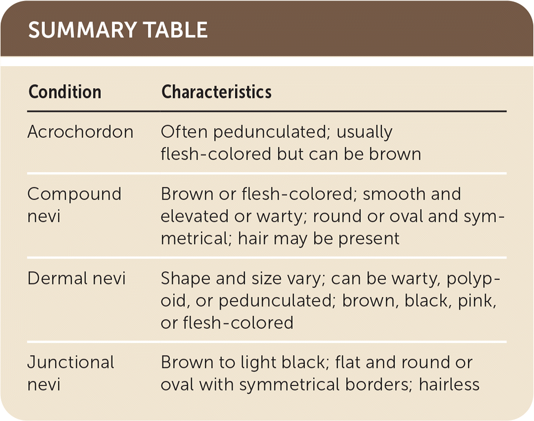
| Condition | Characteristics |
|---|---|
| Acrochordon | Often pedunculated; usually the color of surrounding skin but can be hyperpigmented |
| Compound nevi | Hyperpigmented or the color of surrounding skin; smooth and elevated or warty; round or oval and symmetrical; hair may be present |
| Dermal nevi | Shape and size vary; can be warty, polypoid, or pedunculated; hyperpigmented or pink or appear the same color as surrounding skin |
| Junctional nevi | Hyperpigmented; flat and round or oval with symmetrical borders; hairless |
