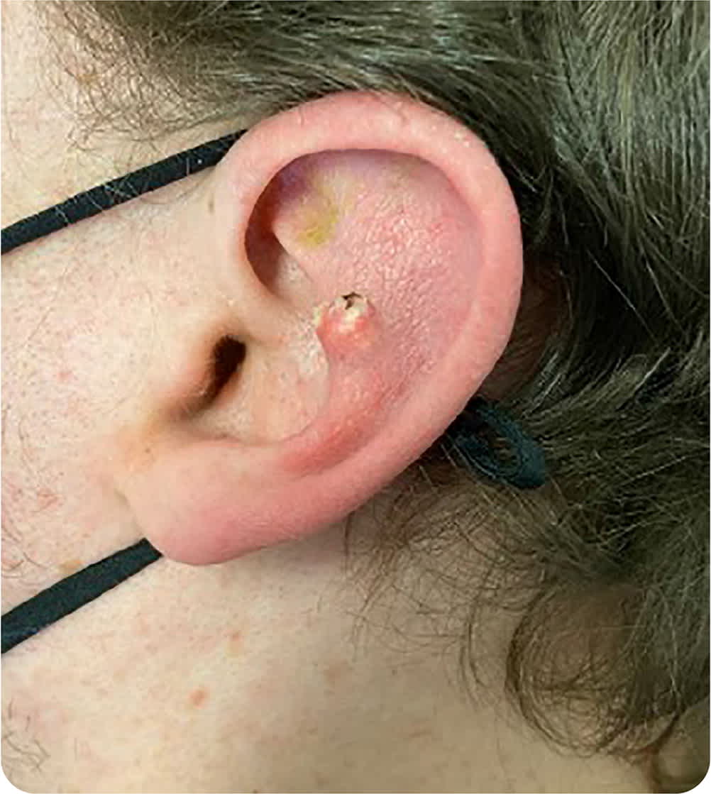
A 34-year-old man presented with a crusted papule on the left ear that had grown slowly over several years. He did not recall any trauma to the area. The lesion was painful, but the patient did not report bleeding or discharge. His medical history included cleft palate repair and gastroesophageal reflux.
On physical examination, a small (less than 1 cm) crusted papule was noted on the left antihelix (Figure 1). The lesion had an erythematous base and was tender to palpation.
FIGURE 1
Question
Based on the patient's history and physical examination findings, which one of the following is the most likely diagnosis?
- A. Basal cell carcinoma.
- B. Chondrodermatitis nodularis helicis.
- C. Gouty tophus.
- D. Squamous cell carcinoma.
- E. Weathering nodule.
Discussion
The answer is C: gouty tophus. A tophus is a deposit of monosodium urate crystals due to hyperuricemia and is pathognomonic for gout. Uric acid tophi may present as hard deposits underneath the skin in and around joints, in the olecranon bursa, or on the pinnae. They may also break through the skin, appearing as chalky white nodules. The diagnosis can be confirmed by aspiration or biopsy of the nodule, which can show needle-shaped, negatively birefringent uric acid crystals under polarized light.1
Removal is not required for small tophi that are not painful and do not affect movement or range of motion. They may shrink over time following appropriate lifestyle changes and diet modification. Medications, including nonsteroidal anti-inflammatory drugs, corticosteroids, and xanthine oxidase inhibitors, can be used prophylactically for gouty tophi. Larger tophi that may affect movement and range of motion should be surgically removed and the joint replaced if indicated.1
Basal cell carcinoma, the most common type of human malignancy, usually occurs on the head and neck. It can present as an enlarging crusted nodule, pearly papule, reddish-pink patch, ulceration, or scar-like area. It usually grows slowly over years, and metastasis is rare.2
Chondrodermatitis nodularis helices is a benign, painful condition that affects the helix or antihelix of the ear. The pathophysiology is unknown but is believed to involve prolonged and excessive pressure on the affected area.3
Squamous cell carcinoma is the second most common skin cancer, with a male to female ratio of 2:1. The incidence increases with age. Lesions are typically nonhealing, bleeding hyperkeratotic nodules or ulcerated plaques, although presentation can vary. The surrounding skin is inflamed and indurated.4 Basal cell and squamous cell carcinomas can be difficult to differentiate clinically without biopsy. Dermoscopy is also useful.
Weathering nodules may be found in the helix and antihelix of the ear. They are most common in older White men and are usually bilateral and occur in multiples. Biopsy shows cartilage with elastic tissue degeneration and a marked absence of inflammatory cells. Weathering nodules may coexist with chondrodermatitis nodularis helices.5
SUMMARY TABLE
| Condition | Characteristics |
|---|---|
| Basal cell carcinoma | Enlarging crusted nodule, pearly papule, reddish-pink patch, ulceration, or scar-like area |
| Chondrodermatitis nodularis helices | Benign and painful condition affecting the helix or antihelix of the ear; possibly caused by prolonged and excessive pressure on affected area |
| Gouty tophi | Associated with chronic hyperuricemia; hard deposits underneath the skin in and around joints, in olecranon bursa, or on pinnae; may break through skin, appearing as chalky white nodules |
| Squamous cell carcinoma | Nonhealing, bleeding hyperkeratotic nodule or ulcerated plaque; surrounding skin inflamed and indurated |
| Weathering nodules | May present in the helix and antihelix of the ear; usually bilateral and multiple; biopsy shows cartilage and elastic tissue degeneration and marked absence of inflammatory cells |
