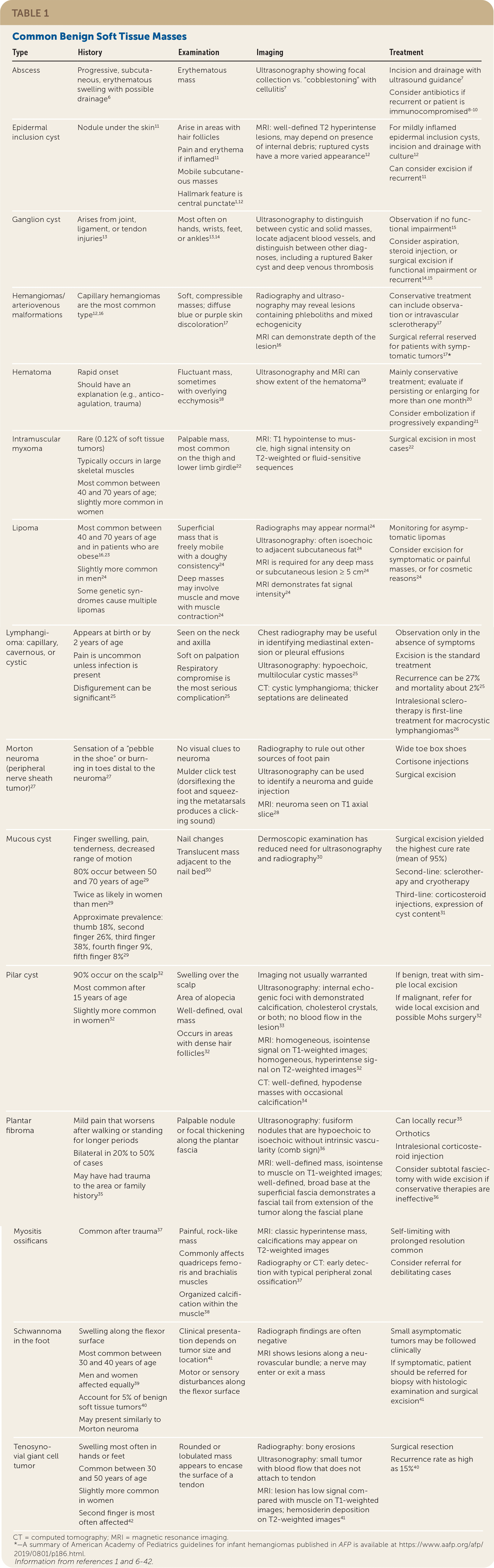
| Type | History | Examination | Imaging | Treatment |
|---|---|---|---|---|
| Abscess | Progressive, subcutaneous, erythematous swelling with possible drainage6 | Erythematous mass | Ultrasonography showing focal collection vs. “cobblestoning” with cellulitis7 | Incision and drainage with ultrasound guidance7 Consider antibiotics if recurrent or patient is immunocompromised8–10 |
| Epidermal inclusion cyst | Nodule under the skin11 | Arise in areas with hair follicles Pain and erythema if inflamed11 Mobile subcutaneous masses Hallmark feature is central punctate1,12 | MRI: well-defined T2 hyperintense lesions, may depend on presence of internal debris; ruptured cysts have a more varied appearance12 | For mildly inflamed epidermal inclusion cysts, incision and drainage with culture12 Can consider excision if recurrent11 |
| Ganglion cyst | Arises from joint, ligament, or tendon injuries13 | Most often on hands, wrists, feet, or ankles13,14 | Ultrasonography to distinguish between cystic and solid masses, locate adjacent blood vessels, and distinguish between other diagnoses, including a ruptured Baker cyst and deep venous thrombosis | Observation if no functional impairment15 Consider aspiration, steroid injection, or surgical excision if functional impairment or recurrent14,15 |
| Hemangiomas/arteriovenous malformations | Capillary hemangiomas are the most common type12,16 | Soft, compressible masses; diffuse blue or purple skin discoloration17 | Radiography and ultrasonography may reveal lesions containing phleboliths and mixed echogenicity MRI can demonstrate depth of the lesion16 | Conservative treatment can include observation or intravascular sclerotherapy17 Surgical referral reserved for patients with symptomatic tumors17* |
| Hematoma | Rapid onset Should have an explanation (e.g., anticoagulation, trauma) | Fluctuant mass, sometimes with overlying ecchymosis18 | Ultrasonography and MRI can show extent of the hematoma19 | Mainly conservative treatment; evaluate if persisting or enlarging for more than one month20 Consider embolization if progressively expanding21 |
| Intramuscular myxoma | Rare (0.12% of soft tissue tumors) Typically occurs in large skeletal muscles Most common between 40 and 70 years of age; slightly more common in women | Palpable mass, most common on the thigh and lower limb girdle22 | MRI: T1 hypointense to muscle, high signal intensity on T2-weighted or fluid-sensitive sequences | Surgical excision in most cases22 |
| Lipoma | Most common between 40 and 70 years of age and in patients who are obese16,23 Slightly more common in men24 Some genetic syndromes cause multiple lipomas | Superficial mass that is freely mobile with a doughy consistency24 Deep masses may involve muscle and move with muscle contraction24 | Radiographs may appear normal24 Ultrasonography: often isoechoic to adjacent subcutaneous fat24 MRI is required for any deep mass or subcutaneous lesion ≥ 5 cm24 MRI demonstrates fat signal intensity 24 | Monitoring for asymptomatic lipomas Consider excision for symptomatic or painful masses, or for cosmetic reasons24 |
| Lymphangioma: capillary, cavernous, or cystic | Appears at birth or by 2 years of age Pain is uncommon unless infection is present Disfigurement can be significant25 | Seen on the neck and axilla Soft on palpation Respiratory compromise is the most serious complication25 | Chest radiography may be useful in identifying mediastinal extension or pleural effusions Ultrasonography: hypoechoic, multilocular cystic masses25 CT: cystic lymphangioma; thicker septations are delineated | Observation only in the absence of symptoms Excision is the standard treatment Recurrence can be 27% and mortality about 2%25 Intralesional sclerotherapy is first-line treatment for macrocystic lymphangiomas26 |
| Morton neuroma (peripheral nerve sheath tumor)27 | Sensation of a “pebble in the shoe” or burning in toes distal to the neuroma27 | No visual clues to neuroma Mulder click test (dorsiflexing the foot and squeezing the metatarsals produces a clicking sound) | Radiography to rule out other sources of foot pain Ultrasonography can be used to identify a neuroma and guide injection MRI: neuroma seen on T1 axial slice28 | Wide toe box shoes Cortisone injections Surgical excision |
| Mucous cyst | Finger swelling, pain, tenderness, decreased range of motion 80% occur between 50 and 70 years of age29 Twice as likely in women than men29 Approximate prevalence: thumb 18%, second finger 26%, third finger 38%, fourth finger 9%, fifth finger 8%29 | Nail changes Translucent mass adjacent to the nail bed30 | Dermoscopic examination has reduced need for ultrasonography and radiography 30 | Surgical excision yielded the highest cure rate (mean of 95%) Second-line: sclerotherapy and cryotherapy Third-line: corticosteroid injections, expression of cyst content31 |
| Pilar cyst | 90% occur on the scalp32 Most common after 15 years of age Slightly more common in women32 | Swelling over the scalp Area of alopecia Well-defined, oval mass Occurs in areas with dense hair follicles32 | Imaging not usually warranted Ultrasonography: internal echogenic foci with demonstrated calcification, cholesterol crystals, or both; no blood flow in the lesion33 MRI: homogeneous, isointense signal on T1-weighted images; homogeneous, hyperintense signal on T2-weighted images32 CT: well-defined, hypodense masses with occasional calcification34 | If benign, treat with simple local excision If malignant, refer for wide local excision and possible Mohs surgery32 |
| Plantar fibroma | Mild pain that worsens after walking or standing for longer periods Bilateral in 20% to 50% of cases May have had trauma to the area or family history35 | Palpable nodule or focal thickening along the plantar fascia | Ultrasonography: fusiform nodules that are hypoechoic to isoechoic without intrinsic vascularity (comb sign)36 MRI: well-defined mass, isointense to muscle on T1-weighted images; well-defined, broad base at the superficial fascia demonstrates a fascial tail from extension of the tumor along the fascial plane | Can locally recur35 Orthotics Intralesional corticosteroid injection Consider subtotal fasciectomy with wide excision if conservative therapies are ineffective36 |
| Myositis ossificans | Common after trauma37 | Painful, rock-like mass Commonly affects quadriceps femoris and brachialis muscles Organized calcification within the muscle38 | MRI: classic hyperintense mass, calcifications may appear on T2-weighted images Radiography or CT: early detection with typical peripheral zonal ossification37 | Self-limiting with prolonged resolution common Consider referral for debilitating cases |
| Schwannoma in the foot | Swelling along the flexor surface Most common between 30 and 40 years of age Men and women affected equally39 Account for 5% of benign soft tissue tumors40 May present similarly to Morton neuroma | Clinical presentation depends on tumor size and location41 Motor or sensory disturbances along the flexor surface | Radiograph findings are often negative MRI shows lesions along a neurovascular bundle; a nerve may enter or exit a mass | Small asymptomatic tumors may be followed clinically If symptomatic, patient should be referred for biopsy with histologic examination and surgical excision41 |
| Tenosynovial giant cell tumor | Swelling most often in hands or feet Common between 30 and 50 years of age Slightly more common in women Second finger is most often affected42 | Rounded or lobulated mass appears to encase the surface of a tendon | Radiography: bony erosions Ultrasonography: small tumor with blood flow that does not attach to tendon MRI: lesion has low signal compared with muscle on T1-weighted images; hemosiderin deposition on T2-weighted images41 | Surgical resection Recurrence rate as high as 15%40 |