A 32-year-old man with well-controlled HIV infection presented with a chief symptom of severe rectal pain that worsened over the previous week. He had receptive anal intercourse 13 days before presentation. He described a burning or tearing sensation inside his rectum and an associated clear discharge.
One week earlier, he had diarrhea for two days and developed a rash that started on his legs and spread to his arms and back. He also had a large lesion on his forehead. Two days previously, he presented to another clinic with subjective fevers and night sweats and was treated with doxycycline and ceftriaxone. He did not have chest pain, shortness of breath, or abdominal pain. He did not have recent travel or known infectious contacts.
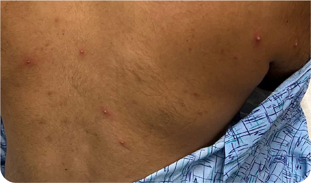
Physical examination revealed diffuse, salmon-colored macules and papules on his left arm, papules and vesicles on his back, and an umbilicated lesion on his forehead (Figure 1 and Figure 2).
FIGURE 1

FIGURE 2
Question
Based on the patient's history and physical examination findings, which one of the following is the most likely diagnosis?
- A. Gonorrhea.
- B. Herpessimplex.
- C. Mpox (monkeypox).
- D. Syphilis.
- E. Ulcerativecolitis.
Discussion
The answer is C: mpox. Rectal pain in men who have sex with men is a common presentation.1 The patient's history of constitutional symptoms (fever and night sweats), rectal pain with discharge following anal intercourse, and rash (including an umbilicated lesion on the forehead) are characteristic of mpox. Patients with mpox may also have other sexually transmitted infections, and diseases such as COVID-19, influenza, and varicella.
HIV infection and men having sex with men are risk factors for mpox. The incubation period after exposure ranges from five to 21 days. Monkeypox is a cytopathic virus with an affinity for epithelial cells. It produces mucosal lesions (enanthema), and then a rash. Skin lesions typically progress through macular (one or two days), papular (one or two days), vesicular (one or two days), and pustular (five days) stages before scabbing (seven to 14 days). Healing may occur in multiple stages in different body regions. Pustular lesions are usually painful, deep-seated, well-circumscribed, and umbilicated.2 The characteristic umbilication of the lesion helps distinguish mpox from other diseases. Clear, bloody, or purulent discharge may occur.
Treatment of mpox is mainly supportive and includes pain management and monitoring for rare but serious complications, including blindness, urinary obstruction, rectal perforation, and lymphadenopathy leading to airway obstruction. Tecovirimat (Tpoxx) provides analgesia and prevents severe disease progression. Debilitating pain and disfiguring scars are possible with mpox. However, the disease is usually self-limited, resolving in two to four weeks.
Gonorrhea is caused by Neisseria gonorrhoeae. It primarily affects the genitourinary tract, leading to anorectal complications such as mucopurulent discharge with tenesmus, pruritus, and bleeding.3 Anoscopy or proctoscopy may reveal yellow, mucous discharge from the anal crypts. Skin lesions in disseminated infections include small, scattered, purpuric macules on the palms, which can evolve into vesicopustules, sometimes with hemorrhage but without umbilication.4
Symptoms of herpes simplex are usually mild but can be moderate to severe. Patients with rectal herpes present with proctitis, anorectal pain and discharge, tenesmus, and rectal bleeding. The acute phase is characterized by difficulty urinating, sacral paresthesias, and temporary fecal incontinence. Severe cases present as perianal erythema and ulcerations manifesting as clusters of red or pink, liquid-filled blisters that do not umbilicate, typically near the site of infection.
In primary syphilis, a chancre (punched-out, painless ulcer) occurs at the site of infection, usually in the mucosa of the mouth, anus, or genitals.5 Syphilitic anal lesions can be painful or painless. Patients may have anal fissures, often with lymphadenopathy. Proctitis, a less common presentation, produces erythematous/hyperemic rectal masses that sometimes ulcerate.6,7 Patients may have swollen infiltrates in the distal rectum or anal canal that can produce a large mass. Positive serology findings are supportive, but definitive diagnosis requires a nucleic acid amplification test for Treponema pallidum DNA using swabs from suspicious ulcers or mucosal areas.8
Ulcerative colitis often causes proctitis with significant rectal pain, discharge, and bleeding. Patients may develop highly inflammatory, friable, neutrophil-filled, sterile lesions.9 When the eruptions are vesiculopustular, they may include central ulceration and crusting similar to mpox.10
SUMMARY TABLE
| Condition | Characteristics |
|---|---|
| Gonorrhea | Anorectal complications such as mucopurulent discharge with tenesmus, pruritus, and bleeding; when disseminated, small, scattered, purpuric macules on the palms can evolve into vesicopustules |
| Herpes simplex | Clusters of red or pink, liquid-filled blisters, typically near the site of infection; proctitis, anorectal pain and discharge, tenesmus, and rectal bleeding may occur |
| Mpox (monkeypox) | Usually painful, firm or rubbery, well-circumscribed, umbilicated lesions; may have rectal pain with clear, bloody, or purulent discharge |
| Syphilis | Hyperemic, sometimes ulcerated, swollen infiltrates in the distal rectum or anal canal; may produce a large rectal mass |
| Ulcerative colitis | Pain and rectal bleeding; inflammatory skin lesions and vesiculopustular, friable, neutrophil-filled lesions are sterile; may have central ulceration and crusting |
