A 51-year-old man presented with a swollen right knee. The swelling had recurred intermittently for 10 years, staying a consistent size over the past several years. It was aspirated 3 years prior, but the swelling returned quickly. The patient had not received imaging or treatment for his knee. He experienced discomfort with kneeling, dressing, and many daily activities, but did not have knee pain. He had no injuries to the area, recent infections, or history of cancer. The patient had worked as a floor tiler for more than 20 years.
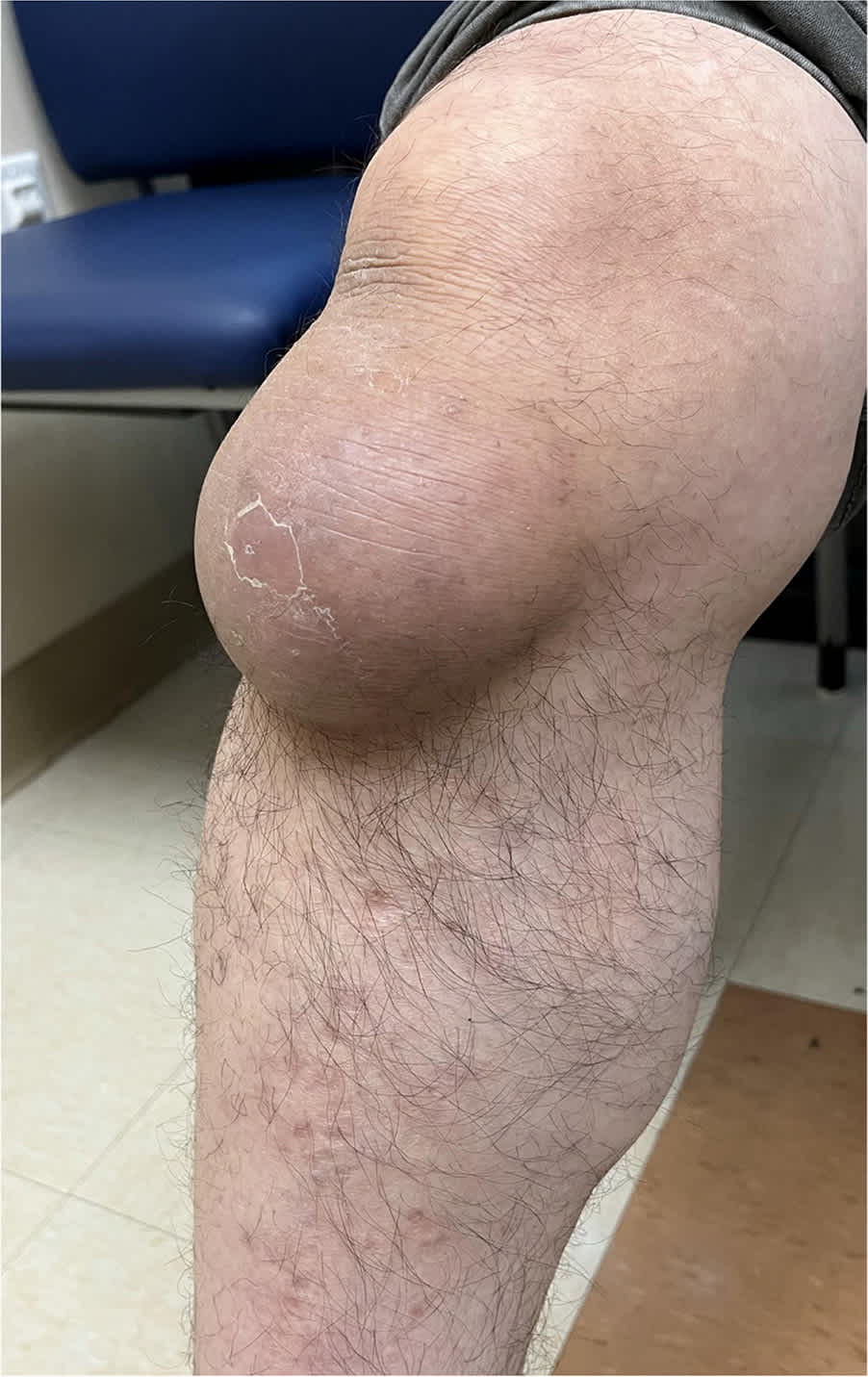
Physical examination revealed that the patient was afebrile and had a 9-cm mass below his right patella (Figure 1). The mass was compressible, fluctuant, cool, and nontender. His patella was grossly normal to palpation superior to the swelling. The skin overlying the lesion was intact, slightly violaceous, and had some desquamation. His active and passive ranges of motion were normal, and the mass did not shift position with flexion or extension of the knee.
FIGURE 1
QUESTION
Based on the patient's history and physical examination, which one of the following is the most likely diagnosis?
- A. Gout.
- B. Osgood-Schlatter disease.
- C. Prepatellar bursitis.
- D. Septic arthritis.
- E. Superficial infrapatellar bursitis.
DISCUSSION
The answer is E: superficial infrapatellar bursitis. This diagnosis is consistent with the patient's presentation of a chronic, nontender fluctuance anterior to the tibial tubercle and inferior to the patella. The condition is associated with an acute injury or chronic knee swelling in occupations that require recurrent kneeling and weight-bearing on the knee.1,2 The location of the superficial infrapatellar bursa between the tibial tubercule and the overlying skin helps identify this condition.3
In the acute phase, treatment of superficial infrapatellar bursitis includes ice, rest, elevation, nonsteroidal anti-inflammatory drugs, and aspiration when the swelling is large or interferes with function.4 Management may also include compression, activity modification, and intrabursal steroid injections. The use of steroids is controversial because of questionable benefit.1,4 Ultrasonography or magnetic resonance imaging can be beneficial in anatomic localization and identifying any related complications.3,4 When there is concern for infection or neoplastic or gouty etiologies, aspiration should be performed.4 Bursectomy can be used in recurrent or complicated cases and in patients who are immunocompromised.4,5
Gout typically presents as swelling, tenderness, and pain in a joint space or bursa. It is most common in men 30 to 50 years of age and in patients with hypertension, diabetes, and obesity. Monosodium urate crystals typically accumulate in distal regions such as the first metatarsophalangeal joint, but gouty arthritis in the knee is not uncommon. Synovial fluid showing monosodium urate crystals with negative birefringence under polarized light is diagnostic for gout.6
Osgood-Schlatter disease presents as anterior knee pain, swelling around the tibial tubercle, and a history of knee flexion stressors.7 It is most prevalent in children and adolescents with a history of repetitive use (eg, sports) due to incomplete skeletal maturity of the tibial tubercle.8 The swelling is more likely to be firm to palpation vs a fluctuant mass. Imaging may show stress or avulsion fracture at the tibial tubercle.1,7
Read the full article
Get immediate access, anytime, anywhere.
Choose a single article, issue, or full-access subscription.
Earn up to 14 CME credits per issue.