The estimated lifetime risk of acquiring a dermatophyte infection is between 10 and 20 percent. Recognition and appropriate treatment of these infections reduces both morbidity and discomfort and lessens the possibility of transmission. Dermatophyte infections are classified according to the affected body site, such as tinea capitis (scalp), tinea bar-bae (beard area), tinea corporis (skin other than bearded area, scalp, groin, hands or feet), tinea cruris (groin, perineum and perineal areas), tinea pedis (feet), tinea manuum (hands) and tinea unguium (nails). To determine the best treatment approach, the physician must consider several factors: (1) the anatomic locations of the infection, (2) the safety, efficacy and cost of treatment options and (3) the likelihood that the patient will comply with treatment. Newer medications in both oral and topical forms, including imidazoles and allylamines, have greatly increased the cure rate for tinea infections. Certain types of tinea may be treated with “pulse” regimens; these innovative therapies lower treatment costs and improve patient compliance.
Superficial fungal infections are among the most common skin diseases,1 affecting millions of people throughout the world.2 These infections, which occur in both healthy and immunocompromised persons, are caused by dermatophytes, yeasts and nondermatophyte molds. Effective treatment can reduce the duration of symptoms in patients with superficial fungal infections.
Dermatophytes, specifically Trichophyton, Epidermophyton and Microsporum species, are responsible for most superficial fungal infections.1,3,4 The estimated lifetime risk of acquiring a dermatophyte infection is between 10 and 20 percent.5
The term “tinea” refers exclusively to dermatophyte infections. Tinea infections are classified according to their anatomic location. (Pityriasis versicolor, sometimes referred to as tinea versicolor, is caused by Malassezia furfur and therefore is not discussed in this article.)
Fungal transmission occurs through direct contact with infected persons, animals, soil or fomites. Depending on their habitat, dermatophytes are described as anthropophilic (human), zoophilic (animal) or geophilic (soil). Anthropophilic dermatophytes are the most common sources of tinea infections, but zoophilic sources should be identified (if possible) and treated to prevent human reinfection.6
The classic presentation of tinea infection, known as “ringworm,” is a lesion with central clearing surrounded by an advancing, red, scaly, elevated border. One or more lesions may appear. Inflammation assists in colonization and may result in vesicles on the border of the affected area. Atopic persons and those infected with zoophilic fungi tend to have more inflammation.
The presentations of tinea infections range from mild scaling and erythema to severe inflammation with bacterial superinfection. The differential diagnosis for suspected tinea infection is listed in Table 1.
TABLE 1 Tinea Infections
| Type | Common causative species | Differential diagnosis |
|---|---|---|
| Tinea capitis | Trichophyton tonsurans | Alopecia areata |
| Microsporum andouinii* | Impetigo | |
| Microsporum canis*† | Pediculosis | |
| Psoriasis | ||
| Seborrhea | ||
| Trichotillomania | ||
| Tinea barbae | Trichophyton verrucosum | Folliculitis |
| Malignant lymphoma | ||
| Sporotrichosis | ||
| Tinea corporis | Trichophyton rubrum | Cutaneous lupus erythematous |
| M. canis* | Drug eruption | |
| T. tonsurans | Eczema | |
| T. verrucosum | Erythema multiforme | |
| Granuloma annulare | ||
| Nummular dermatitis | ||
| Pityriasis rosea | ||
| Pityriasis versicolor* | ||
| Psoriasis | ||
| Secondary syphilis | ||
| Tinea cruris | T. rubrum | Candidal intertrigo |
| Epidermophyton floccosum | Contact dermatitis | |
| Erythrasma* | ||
| Psoriasis | ||
| Seborrhea | ||
| Tinea pedis | T. rubrum | Bacterial or candidal infection |
| Trichophyton mentagrophytes | Contact or atopic dermatitis | |
| var interdigitale | Dyshidrosis | |
| E. floccosum | Eczema | |
| Pitted keratolysis | ||
| Psoriasis | ||
| Tinea manuum | T. rubrum | Same as for tinea pedis |
| Tinea unguium | T. rubrum | Contact dermatitis |
| Trichophyton mentagrophytes | Lichen planus | |
| var mentagrophyte† | Onychodystrophy | |
| Psoriasis |
*—These dermatophytes fluoresce under a Wood's light: Microsporum andouinii and Microsporum canis fluoresce blue-green; Corynebacterium minutissimum, the bacterium that causes erythrasma, fluoresces coral-red; and Malassezia fur-fur, the causative fungus in pityriasis versicolor, fluoresces pale yellow. 6
† —Zoophilic species.
Diagnosis
Wood's Light Examination
Most dermatophytes do not fluoresce. The exceptions are two zoophilic dermatophytes, Microsporum canis and Microsporum andouinii. These minor causes of tinea capitis fluoresce blue-green. Wood's light examination can also help to differentiate erythrasma caused by the bacterium Corynebacterium minutissimum, which fluoresces coral-red, from tinea cruris, which does not fluoresce.
When positive, a Wood's light examination can be helpful in determining the extent of infection, identifying areas for sampling and evaluating treatment response. The examination is also useful for examining the contacts of an infected person.
Microscopy
Microscopic examination is central to the office diagnosis of any tinea infection.7 Material is scraped from an active area of the lesion, placed in a drop of potassium hydroxide solution and examined under a microscope. The examination, which can be done quickly and easily, is highly sensitive and specific for dermatophyte identification.
Microscopy is positive if hyphae are identified in fungal infections and if pseudohyphae or yeast forms are seen in Candida or Pityrosporum infections. A positive examination is sufficient to justify starting treatment,7,8 because species identification does not usually influence treatment choices.7
Culture
Because cultures are both expensive and time-consuming, they are not routinely performed in suspected tinea infections. However, cultures should be obtained when long-term oral drug therapy is being considered, the patient has a recalcitrant infection or the diagnosis is in doubt.
Identification of a specific zoophilic species as the infection source can be helpful in preventing recurrent infection. It is also important to confirm the specific fungus that is causing a nail infection, because the spectrum of activity for oral antifungal agents varies.
Clinical Presentations
Tinea Capitis
Tinea capitis, sometimes called “ringworm of the scalp,” primarily affects school-aged children. It often appears as one or more annular patches of inflammatory or noninflammatory alopecia. Noninflamed areas, characteristic of Trichophyton tonsurans infection, may appear as black dots, which are actually infected hair shafts broken off at the scalp. Sometimes tinea capitis appears only as non-specific dandruff. Microsporum infection presents as gray patches of hairs that are lusterless because of a coating of spores. Inflamed areas usually have scales, pustules and erythema. Some patients develop a localized, boggy, indurated granuloma called a “kerion” (Figure 1). A kerion can cause scarring and permanent hair loss.
FIGURE 1.
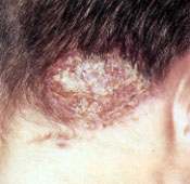
Kerion, a severely inflammatory, boggy, indurated, tumor-like mass that may occur in tinea capitis.
Tinea capitis should be treated with oral therapy. Griseofulvin (Fulvicin PG, Gris-PEG, Grisactin Ultra) is the only oral anti-fungal agent approved by the U.S. Food and Drug Administration for the first-line treatment of tinea capitis. However, itraconazole (Sporanox) and terbinafine (Lamisil) are good alternatives.9–11
Tinea Barbae
Tinea barbae affects the beard area of men who work with animals (Figure 2). It is often accompanied by bacterial folliculitis and inflammation secondary to ingrown hairs. In the treatment of tinea barbae, oral therapy is preferred over topical therapy because the involved hair follicles do not respond well to topical therapy. The agents used to treat tinea capitis are also used to treat tinea barbae.
FIGURE 2.
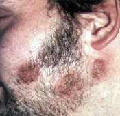
Tinea barbae.
Tinea Corporis
Tinea corporis, also called “ringworm of the body,” often affects children and adults who live in hot, humid climates. The classic presentation of this infection is a circular plaque with a well-demarcated border (Figure 3). Since tinea corporis can be asymptomatic, it can spread rapidly among children in day-care settings.
FIGURE 3.
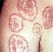
Tinea corporis.
Unless only one or two lesions are present, tinea corporis should be treated orally. Terbinafine and itraconazole are equally effective in treating tinea corporis.12,13 Furthermore, these agents each have a better mycologic cure rate and a lower relapse rate than griseofulvin.14–16 An alternative is fluconazole (Diflucan), which is given orally once a week for up to four consecutive weeks.17,18
Tinea Cruris
Tinea cruris, commonly referred to as “jock itch,” involves the medial aspect of the upper thighs (groin). Unlike yeast infections, tinea cruris generally does not involve the scrotum or the penis (Figure 4). This dermatophyte infection occurs more often in men than in women and rarely affects children.
FIGURE 4.
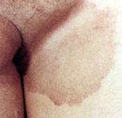
Tinea cruris.
In this infection, erythematous plaques often develop bilaterally. Pruritus is common.
Topical therapy is sufficient in most patients with tinea cruris. If the infection spreads to the lower thighs or buttocks, oral therapy is preferred. Itraconazole and terbinafine are more efficacious than griseofulvin.14,15
Tinea Pedis
Tinea pedis, generally known as “athlete's foot,” is the most common dermatophyte infection. Tinea pedis infection is usually related to sweating and warmth, and use of occlusive footwear. Men between 20 and 40 years of age are most frequently affected. The infection often presents as white, macerated areas in the third or fourth toe webs (Figure 5). It may also present with a classic pattern on the dorsal surface of the foot or as chronic dry, scaly hyperkeratosis of the soles and heels. Because of its distribution, the latter presentation, which is typical of Trichophyton rubrum infection, is sometimes referred to as “moccasin type.”
FIGURE 5.
Tinea pedis involving the toe webs.
Occasionally, tinea pedis may produce acute, highly inflamed, sterile vesicles and pustules at distant sites (arms, chest, sides of fingers). Referred to as the “dermatophytid” or “id” reaction, these vesicles and pustules probably represent an immunologic response to the fungus; they subside when the primary infection is controlled. The “id” reaction can be the only manifestation of an asymptomatic web space maceration.
Tinea pedis is often treated with topical therapy. Since oral agents provide better skin penetration than most topical preparations, oral therapy may be more efficacious in the treatment of hyperkeratotic tinea pedis. Itraconazole, terbinafine and griseofulvin are good choices for oral therapy. Itraconazole and terbinafine are slightly more effective than griseofulvin.14,19 Once-weekly dosing with fluconazole is another option, especially in non-compliant patients.17
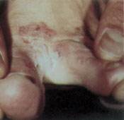
Tinea Manuum
Tinea manuum (Figure 6), a fungal infection of the hands, is less common than tinea pedis. Like tinea pedis, tinea manuum also presents with the classic pattern of erythema and mild scaling on the dorsal aspect of the hands or as a chronic, dry, scaly hyperkeratosis of the palms. When the palms are infected, the feet are also commonly infected. A typical pattern of involvement is either one hand and both feet or both hands and one foot. Treatment options are the same as for tinea pedis.
FIGURE 6.
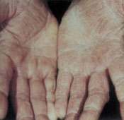
Tinea manuum involving the palms.
Tinea Unguium
Tinea unguium is a dermatophyte infection of the nails. It is a subset of onychomycosis, which includes dermatophyte, nondermatophyte and yeast infections of the nails. Toe-nails are involved more frequently than fingernails. Tinea unguium affects adults more often than children. Risk factors for this fungal infection include increasing age, diabetes, poor venous and lymphatic drainage, ill-fitting shoes and sports participation. However, most infections have no underlying cause. Involvement of the toenail usually implies an extremely resistant infection with a tendency to recur. Chemical or surgical avulsion may be helpful in patients with recalcitrant infection, especially when only a single nail is involved.
The clinical presentation of tinea unguium depends on whether the fungus has invaded the distal, proximal or superficial nail. Distal involvement is the most common presentation. With distal involvement, the affected nail is hyperkeratotic, chalky and dull. The brownish-yellow debris that forms beneath the nail causes the nail to separate from its bed. The nail plate may become brittle, but the nail fold is seldom involved (Figure 7). Although inflammation is uncommon, up to 45 percent of patients experience pain.20 Coexistent tinea manuum or tinea pedis is common.
FIGURE 7.
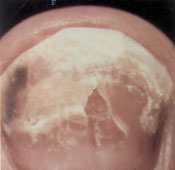
Tinea unguium with distal invasion of the nail.
Tinea unguium requires oral antifungal therapy. Itraconazole, terbinafine and, to a lesser extent, griseofulvin are effective in treating fingernail infections.21–23 Itraconazole or terbinafine are better choices for treatment of toenails.2 The FDA has approved the use of itraconazole “pulse” therapy (i.e., a series of brief medication courses) for the treatment of tinea unguium of the fingernails. The total drug exposure is less with pulse therapy21; therefore, its cost is also lower than traditional treatment. Furthermore, because of the drug-free intervals, patients may be more likely to comply with therapy.
For toenail infections, itraconazole pulse therapy appears to be as effective as daily dosing.24–26 However, the FDA has not approved pulse therapy for treatment of tinea unguium of the toenails.
Terbinafine pulse therapy may also be effective. At this time, however, data are insufficient to recommend pulsed terbinafine therapy.26 Data suggest that fluconazole may be another alternative for treatment of fungal nail infections, especially if yeast is involved. To date, however, few trials of this therapy have been conducted, and only small numbers of patients have been studied.27–29
When itraconazole or terbinafine is used in the treatment of tinea unguium, the nail may not appear clinically cured at the end of therapy. A new nail may require three to 12 months to grow out. Thus, patients should be reminded that the resolution of tinea unguium requires four to six months for fingernails and even longer for toenails.
Treatment Selection
Topical Antifungal Preparations
The topical antifungal agents currently available in the United States are listed in Table 2.30 Although tinea infections are most commonly treated with topical preparations, therapeutic success is limited because of the lengthy duration of treatment, poor patient compliance and high relapse rates at specific body sites.
Factors to consider in selecting an antifungal agent include the route of delivery, efficacy (defined as mycologic and/or clinical cure, and duration of remission), patient compliance and cost. Compared with fungistatic drugs, fungicidal agents are associated with higher cure rates, lower relapse rates and shorter treatment periods.
The clinical state of the lesion also must be considered. Ointment preparations soften thickened, hyperkeratotic lesions. Lotions and solutions prevent maceration in intertriginous areas and hairy areas of the body. They are also appropriate for treating moist, oozy, weepy lesions. Cream formulations are beneficial in the treatment of scaling, non-oozing lesions. Powders alone are less effective in treating existing tinea infections. However, they are helpful as adjunctive agents for reducing moisture and maceration, and they can prevent fungal infections in intertriginous areas.
Topical antifungal agents have been reviewed in American Family Physician.30 Since that time, the first prescription spray, terbinafine 1 percent solution, and a new topical antifungal agent, butenafine 1 percent cream (Mentax), have been approved for use in the treatment of interdigital tinea pedis, tinea corporis and tinea cruris. Butenafine and terbinafine appear to have similar mycologic cure rates, although no direct comparative studies have been performed. Compared with terbinafine, butenafine is effective against fewer organisms. Average wholesale costs for the two drugs are the same.31
Oral Antifungal Agents
Oral therapy is often chosen because of its shorter duration and the potential for greater patient compliance. However, oral agents must be used in the treatment of disease that is extensive, that affects hair and nails, and that does not respond to topical agents.2 Important information about the oral antifungal agents currently available in the United States is presented in Table 3.4,23 Physicians also must be aware of the hematotoxicity and drug interactions of these agents. Drug regimens and medication costs for the treatment of different types of tinea are provided in Table 4.4,9,12–14,16–19,26,32–35 (Ketoconazole [Nizoral] is omitted because it is not a first-line therapy.)
Absorption properties of oral antifungal agents vary. For optimal absorption, griseofulvin should be taken with a fatty meal.23 Ultramicrosize griseofulvin was designed to decrease the need to be taken with food to achieve the best drug absorption. Itraconazole and ketoconazole rely on gastric acidity for optimal absorption. Therefore, patients being treated with these agents should avoid taking antacids, proton pump inhibitors and histamine H2-receptor blockers.
Achlorhydria also decreases the absorption of itraconazole and ketoconazole. If patients drink a cola beverage before they take these drugs, they may acidify their stomach and thereby increase drug absorption by up to 60 percent.36,37 Because the absorption of terbinafine and fluconazole is not influenced by gastric pH, these agents are preferable for use in patients with achlorhydria.22,38 Many oral antifungal agents interact with other medications (Table 3).4,23 Terbinafine has fewer drug interactions because it minimally affects the cytochrome P450 enzyme system.22 Itraconazole, fluconazole and ketoconazole significantly inhibit this system, particularly the subgroup 3A4.21,38,39 Side effects that influence the selection of an oral antifungal agent are also listed in Table 3.4,23 For example, griseofulvin causes side effects in 20 percent of patients.34 This agent most often causes headache or gastrointestinal complaints, but it can also cause rare and more serious adverse reactions, such as toxic epidermal necrolysis and photodermatitis myositis.22,34
Studies of tinea unguium show that itraconazole is better tolerated than griseofulvin.33,40 As with griseofulvin, the most common complaints with itraconazole are headache and gastrointestinal distress. Reversible elevations in liver enzyme levels occur in 0.9 percent of patients treated with itraconazole.41 More serious but rare events include hepatotoxicity, hallucinations, hypokalemia and edema.34,41
Ketoconazole is reserved for the second-line treatment of recalcitrant superficial fungal infections. Of the antifungal agents, this drug has the highest risk for hepatotoxicity, with a reported incidence ranging from one case per 10,00039 to one case per 70,00041 recipients. Patients who are taking ketoconazole should be instructed to immediately report symptoms of hepatotoxicity such as anorexia, nausea and vomiting. Hepatotoxicity appears to be idiosyncratic.42 Risk factors possibly associated with ketoconazole-induced hepatotoxicity include female gender, onychomycosis, alcoholism, ketoconazole therapy lasting more than two weeks and previous griseofulvin treatment.41 Ketoconazole therapy should be discontinued if symptomatic liver inflammation occurs or if the results of liver function tests are three times higher than normal. Liver enzyme levels usually return to normal after ketoconazole is discontinued.42
Ketoconazole is also associated with asymptomatic increases in transaminase levels in 5 to 10 percent of patients.34 More common side effects include gastrointestinal complaints and pruritus.39
The side effects most often associated with fluconazole are rash, headache, gastrointestinal disorders and elevated liver function levels.35,38 Rarely, erythema multiforme can occur in patients treated with this antifungal agent.35
Skin rashes and gastrointestinal side effects are common with terbinafine. Terbinafine has also been associated with Stevens-Johnson syndrome, blood dyscrasias, hepatotoxicity and ocular disturbances, as well as elevated liver enzyme levels in 0.5 percent of recipients.22,41 In addition, some patients have noted losing their sense of taste for up to six weeks.22,41
Adjunctive Topical Corticosteroids
Topical corticosteroids are commonly used as adjuncts to antifungal therapy. These agents are especially beneficial in the initial stages of treatment because they suppress the inflammatory response and provide symptomatic relief. Because of the possibility of fungal proliferation, topical corticosteroids should not be used alone in the treatment of tinea infections.
