
A more recent article on carpal tunnel syndrome is available.
Am Fam Physician. 2003;68(2):265-272
Carpal tunnel syndrome affects approximately 3 percent of adults in the United States. Pain and pares-thesias in the distribution of the median nerve are the classic symptoms. While Tinel's sign and a positive Phalen's maneuver are classic clinical signs of the syndrome, hypalgesia and weak thumb abduction are more predictive of abnormal nerve conduction studies. Conservative treatment options include splinting the wrist in a neutral position and ultrasound therapy. Orally administered corticosteroids can be effective for short-term management (two to four weeks), but local corticosteroid injections may improve symptoms for a longer period. A recent systematic review demonstrated that nonsteroidal anti-inflammatory drugs, pyridoxine, and diuretics are no more effective than placebo in relieving the symptoms of carpal tunnel syndrome. If symptoms are refractory to conservative measures or if nerve conduction studies show severe entrapment, open or endoscopic carpal tunnel release may be necessary. Carpal tunnel syndrome should be treated conservatively in pregnant women because spontaneous postpartum resolution is common.
Carpal tunnel syndrome, the most common focal peripheral neuropathy, results from compression of the median nerve at the wrist.1 The syndrome affects an estimated 3 percent of adult Americans and is approximately three times more common in women than in men.2 High prevalence rates have been reported in persons who perform certain repetitive wrist motions, but the significance of this relationship continues to be challenged. Although 30 percent of frequent computer users complain of hand pares-thesias, only 10 percent meet clinical criteria for carpal tunnel syndrome, and nerve conduction studies are abnormal in only 3.5 percent of these persons.3
Family physicians frequently encounter patients who may have carpal tunnel syndrome. This article reviews the clinical features, diagnosis, and treatment of this relatively common condition.
Clinical Features
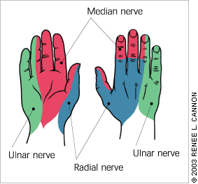
The classic symptoms of carpal tunnel syndrome are pain, numbness, and tingling in the distribution of the median nerve (Figure 1), although numbness in all fingers may be a more common presentation.4 Symptoms are usually worse at night and can awaken patients from sleep. To relieve the symptoms, patients often “flick”their wrist as if shaking down a thermometer (flick sign).
In patients with carpal tunnel syndrome, pain and paresthesias may radiate to the forearm, elbow, and shoulder. Decreased grip strength may result in loss of dexterity, and thenar muscle atrophy may develop if the syndrome is severe. Although one hand typically has more severe symptoms, both hands often are affected.
| Aberrant anatomy |
| Anomalous flexor tendons |
| Congenitally small carpal canal |
| Ganglionic cysts |
| Lipoma |
| Proximal lumbrical muscle insertion |
| Thrombosed artery |
| Infections |
| Lyme disease |
| Mycobacterial infection |
| Septic arthritis |
| Inflammatory conditions |
| Connective tissue disease |
| Gout or pseudogout |
| Nonspecific flexor tenosynovitis* |
| Rheumatoid arthritis |
| Metabolic conditions |
| Acromegaly |
| Amyloidosis |
| Diabetes |
| Hypothyroidism or hyperthyroidism |
| Increased canal volume |
| Congestive heart failure |
| Edema |
| Obesity |
| Pregnancy |
Diagnosis
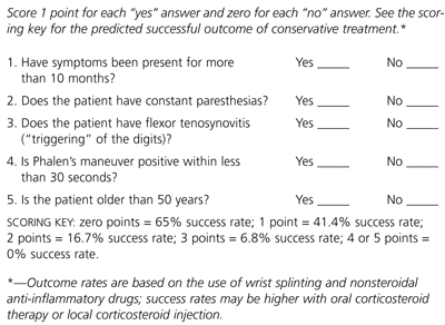
One systematic review7 evaluated the effectiveness of findings from the history and physical examination in predicting positive nerve conduction studies. The most highly predictive findings were symptom location (i.e., a classic or probable pattern marked on hand symptom diagrams), hypalgesia (diminished sensitivity to pain along the palmar aspect of the index finger), and weak thumb abduction.
The principal clinical tests for carpal tunnel syndrome are Phalen's maneuver and Tinel's sign. Phalen's maneuver is positive when flexing the wrist to 90 degrees for one minute elicits symptoms in the median nerve distribution. Tinel's sign is positive when tapping over the carpal tunnel elicits symptoms in the distribution of the median nerve. Sensory findings in carpal tunnel syndrome also may be elicited by two-point discrimination, vibration, and monofilament testing.
Whether carpal tunnel syndrome is a clinical or electrophysiologic diagnosis remains somewhat controversial. In one study of 2,466 persons in a general population,2 354 (14.4 percent) reported pain, numbness, or tingling in the distribution of the median nerve. Nerve conduction studies confirmed the presence of median nerve neuropathy in approximately 45 percent of these symptomatic patients. Interestingly, nerve conduction studies were negative in almost one third of “clinically certain” patients but positive in nearly one third of “clinically uncertain” patients. Of the 125 asymptomatic patients who were examined (control group), 23 (18.4 percent) were found to have median nerve neuropathy on nerve conduction testing.
Treatment
GENERAL MEASURES
Patients with carpal tunnel syndrome should avoid repetitive wrist and hand motions that may exacerbate symptoms or make symptom relief difficult to achieve. If possible, they should not use vibratory tools (e.g., jackhammers, floor sanders), because the motion of these tools can make their symptoms worse.6
Ergonomic measures to relieve symptoms depend on the motion that needs to be minimized. Patients who work on computers, for example, may benefit from improved wrist positioning or the use of wrist supports, although the latter is controversial. Wrist splints may be helpful for patients in other professions that require repetitive wrist motion.
In addition to wrist splinting, conservative treatments include oral corticosteroid therapy and local corticosteroid injections. Approximately 80 percent of patients with carpal tunnel syndrome initially respond to conservative treatment; however, symptoms recur in 80 percent of these patients after one year.9
WRIST SPLINTS
Splinting the wrist at a neutral angle helps to decrease repetitive flexion and rotation, thereby relieving mild soft tissue swelling or tenosynovitis. Splinting is probably most effective when it is applied within three months of the onset of symptoms.11
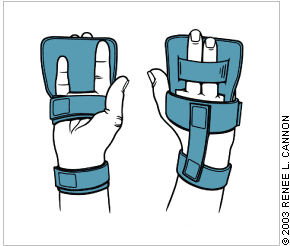
The optimal splinting regimen depends on the patient's symptoms and preferences. Nightly splint use is recommended to prevent prolonged wrist flexion or extension.12 When worn at night for four weeks, a specially designed wrist brace was found to be more effective than no treatment in relieving the symptoms of carpal tunnel syndrome (Figure 3).13 This brace has not yet been compared with traditional splints.
Some patients choose to wear a wrist splint all of the time. Compared with nighttime-only splint use, full-time use has been shown to provide greater improvement of symptoms and electrophysiologic measures; however, compliance with full-time use is more difficult.14 [Evidence level B, uncontrolled trial]
ORAL MEDICATIONS
Diuretics, nonsteroidal anti-inflammatory drugs (NSAIDs), pyridoxine (vitamin B6), and orally administered corticosteroids have been used with varying degrees of success in patients with carpal tunnel syndrome. Unfortunately, few high-quality trials have evaluated these treatments.
A recent systematic review15 of conservative treatments for carpal tunnel syndrome found 14 randomized controlled trials (RCTs), including five trials of oral medications (Table 2).15–20 The authors concluded that NSAIDs, diuretics, and pyridoxine are no more effective than placebo in relieving the symptoms of carpal tunnel syndrome.15 [Evidence level A, systematic review of RCTs] Nevertheless, some investigators21 recommend NSAID therapy as an adjunct to wrist splinting and ergonomic adjustments in patients with mild to moderate carpal tunnel syndrome.
Orally administered corticosteroids have been shown to be more effective than NSAIDs or diuretics in the short-term treatment of carpal tunnel syndrome. The optimal corticosteroid dosage remains to be determined. In one prospective, randomized, double-blind, placebo-controlled trial (73 patients), global symptom scores for carpal tunnel syndrome were significantly improved at two weeks and four weeks in patients randomized to receive prednisolone in a dosage of 20 mg per day for two weeks, followed by 10 mg per day for two weeks.17 [Evidence level B, lower quality RCT] No major adverse effects were noted. The study also found that NSAIDs and diuretics conferred no greater benefit than placebo.
LOCAL INJECTION
Combined injection of a corticosteroid and a local anesthetic into or proximal to the carpal tunnel can be used in patients with mild to moderate carpal tunnel syndrome. Such injections can be diagnostic as well as therapeutic. While most studies evaluating local injection have been retrospective or uncontrolled, two recent systematic reviews of RCTs concluded that local corticosteroid injection provides greater clinical improvement at one month compared with placebo.15,22 [References 15 and 22—Evidence level A, systematic reviews of RCTs]
A randomized, double-blind trial23 compared oral corticosteroid therapy and local corticosteroid injection in 60 patients with electrophysiologically confirmed carpal tunnel syndrome. In this study, 30 patients were treated with local injection of 15 mg of methylprednisolone plus oral placebo; the other 30 patients received 25 mg of orally administered prednisolone for 10 days plus a saline injection. Mean global symptom scores for the two groups did not differ significantly at two weeks. At eight and 12 weeks, however, significant improvement was evident only in the methylprednisolone injection group.
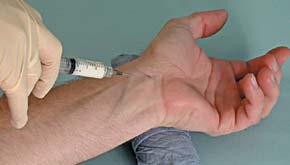
Direct injection into the carpal tunnel by either method carries the potential for needle injury to the median nerve, intratendinous injection and tendon rupture, or dysesthesias (secondary to intrafascicular injection) that may persist for months.26 An alternative approach is to place the injection proximal to the carpal tunnel, rather than directly in it (Figure 5). This approach lowers the risk of damage to the median nerve and theoretically treats concomitant swelling at the volar side of the forearm. With any method, injection of corticosteroid into the median nerve must be avoided.
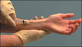
In one double-blind, placebo-controlled trial,27 60 patients older than 18 years with electrophysiologically confirmed carpal tunnel syndrome and symptoms for longer than three months were randomized to receive 10 mg of lignocaine (lidocaine) or 10 mg of lignocaine plus 40 mg of methylprednisolone. In all patients, the injection was administered proximal to the carpal tunnel. Improvement was defined as no symptoms or minor symptoms requiring no further treatment. At one month, symptoms were improved in 23 (77 percent) of the 30 patients in the cortico-steroid-treated group compared with six (20 percent) of the 30 patients in the control group (P<.001); the number needed to treat was 1.8.27 [Evidence level A, RCT] No side effects were reported. Superior symptom relief for corticosteroid injection compared with placebo (lignocaine only) injection was not adequately demonstrated beyond one month of follow-up. All injections in the study were performed by one neurologist, which may limit the reproducibility of the results.
Splinting is generally recommended after local corticosteroid injection.5,25 If the first injection is successful, a repeat injection can be considered after a few months.9 Surgery should be considered if a patient needs more than two injections.9 While a poor response to local corticosteroid injection does not predict a poor surgical result, a favorable response has been associated with a satisfactory surgical outcome.5
ULTRASOUND THERAPY
Ultrasound therapy may be beneficial in the longer term management of carpal tunnel syndrome. A double-blind, randomized trial28 found that compared with “sham ultrasound” treatment (control), 20 sessions of carpal tunnel ultrasound therapy administered over approximately seven weeks resulted in significantly greater improvement of symptoms at two weeks, seven weeks, and six months. However, a smaller study showed no benefit for this treatment.29 More studies are needed to confirm the usefulness of ultrasound therapy for carpal tunnel syndrome.
Additional randomized trials comparing conservative therapies for carpal tunnel syndrome would be useful in helping physicians select appropriate treatments for individual patients.
SURGERY
Carpal tunnel release surgery should be considered in patients with symptoms that do not respond to conservative measures and in patients with severe nerve entrapment as evidenced by nerve conduction studies, thenar atrophy, or motor weakness. It is important to note that surgery may be effective even if a patient has normal nerve conduction studies.30,31
Carpal tunnel release surgery is an out-patient procedure that is performed using regional anesthesia. The traditional surgical approach uses a long palmar curvilinear incision to facilitate division of the transverse carpal ligament and its overlying structures. Endoscopic carpal tunnel release is a newer procedure that allows division of the transverse carpal ligament with the overlying structures left intact. Use of this procedure purportedly lessens scar formation and allows an earlier return to work and activities of daily living. The wrist is generally splinted for three to four weeks after surgery.
One systematic review34 found equal effectiveness for endoscopic and open surgical techniques in relieving the symptoms of carpal tunnel syndrome, but conflicting evidence on whether endoscopic surgery allows an earlier return to work or other activities. Four studies in the review demonstrated earlier return to activity after endoscopic carpal tunnel release, whereas three studies demonstrated no difference between the techniques.
Complications of surgery include injury to the palmar cutaneous or recurrent motor branch of the median nerve, hypertrophic scarring, laceration of the superficial palmar arch, and tendon adhesion.25 Postoperative infection, hematoma, arterial injury, stiffness, and reflex sympathetic dystrophy are other possible complications.21 If carpal tunnel release is incomplete, symptoms may recur.
Incorrect or incomplete diagnosis may be a factor in explaining unsuccessful carpal tunnel release surgery. The strongest predictors of an unfavorable surgical outcome are poor scores on patient-reported measures of upper extremity function and mental health status, compensation action connected to carpal tunnel syndrome that involves an attorney, and alcohol use.32
PREGNANCY
Alterations in fluid balance may predispose some pregnant women to develop carpal tunnel syndrome. Symptoms are typically bilateral and first noted during the third trimester. Conservative measures are appropriate, because symptoms resolve after delivery in most women with pregnancy-related carpal tunnel syndrome.35