Cryosurgery is a highly effective treatment for a broad range of benign skin problems. With appropriate instruction and supervised experience, family physicians can master the technique quickly. Cryosurgery is best suited for use in patients with light skin and for treatment of lesions in most non–hair-bearing areas of the body. Spray methods include the timed spot freeze technique, the rotary or spiral pattern, and the paintbrush method. Benign skin lesions that are suitable for freezing include actinic keratosis, solar lentigo, seborrheic keratosis, viral wart, molluscum contagiosum, and dermatofibroma. Cryosurgery requires little time and fits easily into the physician’s office schedule. Advantages of this treatment include a short preparation time, low risk of infection, and minimal wound care. In addition, cryosurgery requires no expensive supplies or injectable anesthesia, and the patient does not have to return for suture removal. Potential side effects include bleeding, blister formation, headache, hair loss, and hypopigmentation, but rarely scarring. Skin lesions often can be treated in a single session, although some require several treatments.
Cryosurgery has been used to treat skin lesions for approximately 100 years. The first cryogens were liquid air1 and compressed carbon dioxide snow.2 Liquid nitrogen became available in the 1940s and currently is the most widely used cryogen.
Over the past 50 years, much experience has been gained in the use of cryosurgery to treat skin lesions.3 The cotton-tipped dipstick method of liquid nitrogen application has been popular in the management of common benign lesions (Figure 1, left). However, this method is being supplanted by liquid nitrogen spray techniques (Figure 1, center). Liquid nitrogen spray equipment (Figure 2) is easy to use, and similar techniques can be employed to manage benign, premalignant, and malignant lesions.
FIGURE 1.
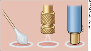
Cryosurgery devices. (Left) Cotton-tip applicator. (Center) Liquid nitrogen spray. (Right) Cryoprobe.
Mechanism of Action
Liquid nitrogen, which boils at −196°C (−320.8°F), is the most effective cryogen for clinical use. It is particularly useful in the treatment of malignant lesions. Temperatures of −25°C to −50°C (−13°F to −58°F) can be achieved within 30 seconds if a sufficient amount of liquid nitrogen is applied by spray or probe. Generally, destruction of benign lesions requires temperatures of −20°C to −30°C (−4°F to −22°F). Effective removal of malignant tissue often requires temperatures of −40°C (−40°F) to −50°C.
Irreversible damage in treated tissue occurs because of intracellular ice formation. The degree of damage depends on the rate of cooling and the minimum temperature achieved. Inflammation develops during the 24 hours after treatment, further contributing to destruction of the lesion through immunologically mediated mechanisms.
FIGURE 2.
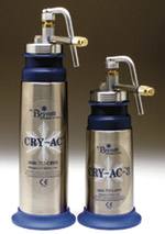
Liquid nitrogen spray equipment. (Left) Cryogun. (Right) Mini Cryogun.
Slow thaw times and repeat freeze-thaw cycles produce more tissue injury than a single freeze and thaw. Usually, several minutes are allowed between repeat freeze-thaw cycles. Repeat freeze-thaw cycles generally are employed only in the treatment of malignancy.
Continuous freezing at one location for more than 30 seconds beyond when an adequate freeze ball is achieved around the target area can result in disruption of the collagen matrix of the skin and possible scarring.
FIGURE 3.
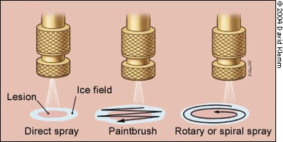
Liquid nitrogen spray patterns.
Mild freezing leads to dermoepidermal separation, which is useful in treating benign epidermal lesions. The more sensitive cells in the epidermis are destroyed while the dermis is left intact. Treatment may be complicated by an element of hypopigmentation, but studies and clinical experience indicate that repigmentation often occurs over several months because of undamaged melanocytes within hair follicles or the migration of melanocytes from the edge of the frozen zone.4 However, the predictability of repigmentation in individual patients is uncertain.
Methods of Treatment
The dose of liquid nitrogen and the choice of delivery method depend on the size, tissue type, and depth of the lesion. The area of the body on which the lesion is located and the required depth of freeze also should be considered. Additional patient factors to consider include the thickness of the epidermis and underlying structures, the water content of the skin, and local blood flow.
Liquid nitrogen spray methods for lesions of different sizes include the timed spot freeze or direct spray technique, the rotary or spiral pattern, and the paintbrush method (Figure 3).
TIMED SPOT FREEZE TECHNIQUE
The timed spot freeze technique allows greater standardization of liquid nitrogen delivery. It may be the most appropriate method for physicians who are learning to perform cryosurgery. Use of this technique maximizes the ability to destroy a lesion with minimal morbidity. The freezing time is adjusted according to variables such as skin thickness, vascularity, tissue type, and lesion characteristics.
Timed spot freezing is performed with a small spray gun that typically holds 300 to 500 mL of liquid nitrogen. Nozzle sizes range from A through F, with F representing the smallest aperture. Nozzle sizes B and C are suitable for the treatment of most benign and malignant lesions; they are the apertures most frequently noted in case reports.
For the standard spot freeze technique, the nozzle of the spray gun is positioned 1 to 1.5 cm from the skin surface and aimed at the center of the target lesion (Figure 4). The spray gun trigger is depressed, and liquid nitrogen is sprayed until an ice field (or ice ball) encompasses the lesion and the desired margin (Figure 5). The designated ice field may need to be delineated in advance with a skin marker pen, because freezing may blur pretreatment lesion margins.
The margin size depends primarily on the thickness of the lesion and whether the lesion is benign or malignant. Margins for most benign lesions can extend as little as 1 to 2 mm beyond the visible pathologic border. Premalignant lesions need margins of 2 to 3 mm, while malignant lesions require margins of 5 mm of clinically normal skin to ensure adequate removal. These margin sizes allow enough depth of freeze to ensure temperatures of −50°C to a depth of 4 to 5 mm.
Once the ice field has filled the specified margin, the spray needs to be maintained, with the spray canister trigger pressure and, thus, the liquid nitrogen spray flow adjusted to keep the target field frozen for an adequate time. This time may vary from five to 30 seconds beyond the initial time for formation of the ice field. If more than one freeze-thaw cycle is required for lesion destruction, complete thawing should be allowed before the next cycle (usually two to three minutes).
The timed spot freeze technique achieves temperatures that are adequate for tissue destruction in an ice field up to 2 cm in diameter. The best approach for lesions larger than 2 cm (including an adequate margin) is to use overlapping treatment fields. A detailed discussion of this approach is beyond the scope of this article.
OTHER TECHNIQUES
FIGURE 4.
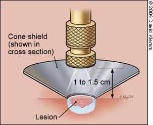
Liquid nitrogen spray application using a timed spot freeze technique and an open cone shield to direct the liquid nitrogen (note that an otoscope tip can be used with similar effect). The spray nozzle is positioned approximately 1 to 1.5 cm above the target lesion.
FIGURE 5.
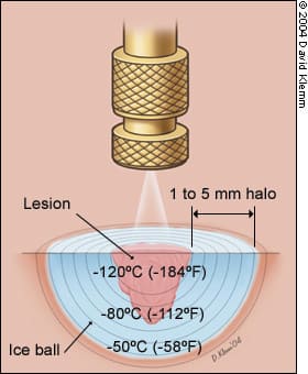
Timed spot freeze technique used to treat a malignancy (possibly a small basal cell cancer), demonstrating freeze ball formation and the 5-mm treatment margins necessary to achieve a temperature of −50ºC (−58 ºF) and, thus, the required depth of 4 to 5 mm.
Variations on the open spray technique include the rotary or spiral pattern and the paintbrush method. These techniques can be useful for treating larger benign lesions. They are not well standardized for ensuring the temperatures that are required for the destruction of malignant lesions.5,6
CRYOPROBES
While the open spray technique can be used for the most easily accessible lesions, a cryoprobe (Figure 1, right) attached to the liquid nitrogen spray gun can provide added versatility, depending on the site and type of the lesion. Various sizes and types of cryoprobes are available (Figure 6). The cryoprobe is applied directly to the lesions. A gel interface medium often is used between the probe and the skin surface.
Cryoprobes frequently are used in the treatment of smaller facial lesions (e.g., on the eyelids), where scatter of liquid nitrogen is undesirable. Probes also are useful in the management of vascular lesions, where the pressure of the probe can be used to remove blood from the tissues thus allowing more adequate treatment (Figure 6).
Treatment of Benign Cutaneous Lesions
Most benign skin lesions can be treated successfully with any of several treatment modalities (excision, cryosurgery, electrodesiccation curettage). However, cosmesis, cost, and patient convenience may make one treatment modality more desirable than another. Patients should be informed about all treatment options and should be allowed to choose from the reasonable alternatives.
FIGURE 6.
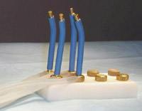
Mini Probe. The probe comes in different sizes (1 to 6 mm), with spray nozzles of variable aperture size.
For the conditions discussed in this article, cryosurgery has advantages over the other modalities. Preparation time is short, and treatment requires no expensive supplies or injectable anesthesia. In addition, the risk of infection is low, wound care is minimal, and suture removal is not needed.7
Correct clinical diagnosis and lesion selection are as critical as the timing of the liquid nitrogen spray in producing a favorable outcome. Depending on the type of lesion, standard technique may need to be varied. Table 1 summarizes cryosurgery techniques for a variety of benign skin lesions.6,8–10
SUN-DAMAGED SKIN
Sun-damaged skin and related lesions are highly amenable to intervention with cryosurgical treatment. Localized small actinic keratosis, which is one of the most common solar-related skin abnormalities, usually requires only five to seven seconds of freeze time beyond initial appearance of a halo around the target lesion. Treatment requires only one freeze-thaw cycle and a margin of less than 1 mm.
TABLE 1 Recommended Cryosurgical Techniques
| Type | Technique | Freeze time (seconds)* | Number of FTCs | Margin (mm) | Number of treatment sessions | Interval (weeks)† |
|---|---|---|---|---|---|---|
| Actinic keratosis | OS | 5 | 1 | 1 | 1 | |
| Cherry angioma | P | 10 | 1 | < 1 | 1 | |
| Common warts | OS | 10 | 1 | 2 | 3 | 4 |
| Cutaneous horn | OS | 10 to 15 | 1 | 2 | 1 | |
| Dermatofibroma | P/OS | 20 to 30 | 1 | 2 | 2 | 8 |
| Hypertrophic scar | OS/P | 20 | 1 | 2 | 1 | |
| Ingrown toenail‡ | OS | 20 | 1 | 2 | 2 | 8 |
| Keloid | OS/P | 20 to 30 | 1 | 2 | 3 | 8 |
| Myxoid cyst | OS/P | 20 | 1 | < 1 | 1 | |
| Oral mucocele | P | 10 | 1 | < 1 | 1 | |
| Pyogenic granuloma | OS | 15 | 1 | < 1 | 1 | |
| Sebaceous hyperplasia | P | 10 | 1 | < 1 | 3 | 4 |
| Skin tags | F/OS | 5 | 1 | 2 | 1 | |
| Solar lentigo | OS | 5 | 1 | < 1 | 1 |
FTC = freeze-thaw cycle; OS = open spray; P = cryoprobe; F = forceps. *—Interval after the initial freeze ball or halo forms, not the total spray time.
†—Interval between repeat treatment sessions.
‡—Target is inflamed granulation tissue, and treatment is helpful in selected cases.
SEBORRHEIC KERATOSIS
Seborrheic keratosis, the most common benign neoplasm, is best treated with cryosurgery or shave excision/curettage. Cryosurgery is especially effective in patients with multiple lesions.11 Thin, flat lesions usually require only one five- to 10-second freeze-thaw cycle; larger, thicker lesions may need longer treatments times or, occasionally, two freeze-thaw cycles.
In treating seborrheic keratosis, the physician should consider the potential for hair loss in treated areas when choosing a therapeutic modality. The other major side effect of cryosurgery for this lesion is hypopigmentation. This side effect is more likely to occur in patients with dark skin.
VIRAL SKIN INFECTIONS
Warts that are resistant to over-the-counter topical agents commonly are treated with cryosurgery. However, response is variable and often depends on the size of the wart and the degree of hyperkeratosis. Several treatment sessions typically are required, and the overall success rate is approximately 75 percent.12,13
To avoid hypopigmentation, small flat warts may be treated with a light spray technique. Digital warts respond favorably to the timed spot freeze spray technique. Deep plantar or palmar warts present challenges, because pain may limit the patient’s tolerance of freezing. However, favorable cure rates have been reported for initial pretreatment with keratolytics (salicylic acid).13 The wart then can be shaved, and the base can be frozen with or without multiple freeze-thaw cycles.14
Cryosurgery has been found to be effective in the management of condyloma acuminatum, particularly when treatment with podophyllin (Podocon-25) has failed or the lesion is located in an area where use of this agent is undesirable.
Molluscum contagiosum, a common dermatologic problem in younger persons, is caused by a poxvirus. This lesion is amenable to cryosurgery, if indicated.15 Applying liquid nitrogen spray for a few seconds until the surface of the umbilicated papule turns white usually is adequate.
TABLE 2 Contraindications to Cryosurgery
| Absolute contraindications | Relative contraindications |
| Lesion for which tissue pathology is required* Lesion located in an area with compromised circulation Melanoma Patient unable to accept possibility of pigmentary changes Proven sensitivity or adverse reaction to cryosurgery Sclerosing basal cell carcinoma or recurrent basal cell or squamous cell carcinoma, particularly when located in a high-risk area (e.g., temple, nasolabial fold) | Cold intolerance Cold urticaria Collagen disease or autoimmune disease Concurrent treatment with immunosuppressive drugs Cryoglobulinemia Heavily pigmented skin Lesions located in pretibial areas, eyelid margins, nasolabial fold, ala nasi, and hair-bearing areas Multiple myeloma Pyoderma gangrenosum Raynaud’s disease |
*—Biopsy must be performed before cryosurgery is considered.
DERMATOFIBROMA
Open spray or cryoprobe techniques may be used to improve the cosmetic appearance of dermatofibromas. Surgical excision of these deep, asymptomatic skin nodules may result in hypertrophic scar formation, because the fibrous lesions are thought to arise from skin microtrauma.
TABLE 3 Complications and Side Effects of Cryosurgery
| Acute |
| Bleeding at the freeze site |
| Blister formation |
| Edema |
| Headache (after treatment of facial lesions) |
| Pain |
| Syncope (vasovagal; rare) |
| Delayed |
| Bleeding |
| Excess granulation tissue formation (rare) |
| Infection (rare) |
| Protracted or permanent |
| Atrophy (rare) |
| Hair and hair follicle loss |
| Hypopigmentation |
| Protracted but temporary |
| Alteration of sensation |
| Hyperpigmentation |
| Hypertrophic scarring |
| Milia |
A single 20- to 30-second freeze-thaw cycle is advised, and a 1- to 2-mm margin should be obtained. Retreatment in eight weeks may be necessary. Significant clinical improvement, including visible flattening of raised dermatofibromas and lightening of pigmentation, has been reported in 80 to 90 percent of patients.16
Contraindications
The relatively few contraindications to cryosurgery generally are related to concomitant illnesses in which excess reactions to cold may occur or delayed healing may be anticipated (Table 2). Some relative contraindications may make alternative treatment modalities more suitable. Physicians often do not perform cryosurgery in the pretibial areas, especially in elderly patients, because of slow wound healing.
Complications
Common complications and side effects of cryosurgery are listed in Table 3. Skin discomfort, generally a burning sensation, occurs with cryosurgery, but intensity is variable. The most sensitive areas are the fingertips, ears, and temples. Freezing of lesions on the forehead or temple may produce headaches. Treatment in hair-bearing areas can result in permanent hair loss.
Hypopigmentation is common, especially with longer freeze times, but is less noticeable in light-skinned patients and improves within several months. Hypopigmentation is caused by the greater sensitivity of melanocytes to freezing, a situation that can be used to advantage in the treatment of dermatofibromas, which frequently have some mild overlying hyperpigmentation. Feathering of the freeze margin (lighter freeze area) often results in a better transition of pigmentary changes.
Freezing for less than 30 seconds beyond initial freeze ball formation does not result in scarring because of the preservation of fibroblasts and the collagen layer of the dermis, which allows for in-migration of the cellular components in the healing process and normal integrity of the skin layers.
Although rare and usually temporary, sensory nerve damage has been reported occasionally in large case series.17,18 It may take 12 to 18 months for sensation to return.
Strength of Recommendations
| Key clinical recommendation | Strength of recommendation | Reference |
|---|---|---|
| In the treatment of deep plantar or palmar warts, favorable cure rates have been reported for initial pretreatment with keratolytics (salicylic acid). | A | 13 |
| Molluscum contagiosum, a common dermatologic problem in younger persons, is caused by a poxvirus. This lesion is amenable to cryosurgery, if indicated. | C | 15 |
