
Am Fam Physician. 2003;68(10):1955-1961
A more recent article on pigmentation disorders is available.
This is part I of a two-part article on hyperpigmentation in adults. Part II, “Melanoma, Seborrheic Keratoses, Acanthosis Nigricans, Melasma, Diabetic Dermopathy, Tinea Versicolor, and Postinflammatory Hyperpigmentation,” appears in this issue on page 1963.
The cause of hyperpigmentation usually is traced to the activity and presence of melanocytes. Café au lait macules may be solitary benign findings or may indicate the presence of neurofibromatosis with its associated complications. Diffuse hyperpigmentation should prompt a search for offending medications or systemic diseases such as hemochromatosis, hyperthyroidism, and Addison's disease. In these instances, the hyperpigmentation may be ameliorated by discontinuing offending medications, performing serial phlebotomy in patients with hemochromatosis, instituting cause-specific treatments in patients with hyperthyroidism, and replacing deficient glucocorticoids and mineralocorticoids in patients with Addison's disease. Cosmetic treatment with bleaching agents or lasers can be used to decrease pigmentation of ephelides (freckles) and lentigines.
Hyperpigmentation may be the sign of a benign or relatively easily treated condition, or it may indicate the presence of a life-threatening condition such as melanoma. This two-part article presents a suggested approach to adult patients with increased pigmentation and reviews various underlying causes.
Melanocytes and Pigmentation
Melanocytes are derived embryonically from neural crest cells that migrate into the basal layer of the epidermis. In the skin, melanocytes continuously produce melanosomes—organelles that are transferred to keratinocytes. The melanosomes convert tyrosine to melanin, giving skin its color. Under the stimulus of hormones or irritation, the production of melanosomes increases, leading to hyperpigmentation. In response to sun exposure or idiopathically in some disorders, hyperplasia of melanocytes occurs and causes hyperpigmentation.1
Diagnostic Approach
A simple way to approach hyperpigmentation is to consider whether the increase in color is caused by an increase of melanin, an increase in melanocytes, or the deposition of another substance that adds color to the skin (Figure 1). Selected disorders and their characteristics are discussed in the following sections and summarized in Table 1; other disorders are discussed and summarized in part II of this article.
| Disorder | Appearance | Age of onset | Hormone or medication related | Sun or other exposure involved | Associated systemic complications | Treatment |
|---|---|---|---|---|---|---|
| Café au lait macules | Multiple macules, 0.2 to 30 cm in diameter, with smooth or irregular, but discrete borders | Congenital or in childhood | Not related | Not involved | May indicate neurofibromatosis (see Table 2) | Treat skin lesions for cosmesis; use laser or surgical procedure if mass effect. |
| Addison's disease | Diffuse hyperpigmentation with “muddy” appearance, especially in sun-exposed areas, perineum, axillae, areolae, palms, and soles | Usually adulthood | Elevated adrenocorticotropic hormone and melanocyte-stimulating hormone levels | More prominent in sun-exposed areas | Blood pressure instability, fatigue, anorexia, depression | Use hydrocortisone and fludrocortisone (Florinef). |
| Hemochromatosis | Diffuse slate-gray or bronze hyperpigmentation | Adulthood | Not related | Not involved | Multiorgan dysfunction caused by iron deposition | Use phlebotomy to decrease iron load and address end-organ damage. |
| Ephelides (freckles) | Multiple red, tan, or brown macules, 1 to 3 mm in diameter, on sun-exposed areas | Childhood through adulthood | Not related | Increased number and pigmentation | None | Treat for cosmesis; avoid sun exposure; use sunscreen, bleaching or peeling agents, or laser. |
| Lentigines | Multiple tan, brown, or black macules, 2 to 20 mm in diameter, on sun-exposed areas | Childhood through adulthood | Not related | Increased in sun-exposed areas | If present on mucosa, consider Peutz-Jeghers syndrome (gastrointestinal polyposis and increased cancer risk) | Treat for cosmesis; avoid sun exposure; use sunscreen, bleaching or peeling agents, or laser therapy. |
| Photoallergic/phototoxic reaction | Diffuse inflammation followed by hyperpigmentation in sun-exposed areas | Any age | Not related | Combination of sun exposure and an offending medication, plant, or chemical | None | Treat for cosmesis;- avoid sun exposure and the offending agent; may use bleaching agents, laser therapy, or dermabrasion. |
A directed history and physical examination offer clues to the underlying cause of hyperpigmentation. The history should address the time of onset of the lesion, because some disorders (e.g., neurofibromatosis) are congenital, while others develop in childhood (e.g., ephelides) or during pregnancy (e.g., melasma). Systemic symptoms may indicate the presence of hyperthyroidism, Addison's disease, or diabetes-related disorders. A review of medication use, supplement use, and exposure to plants and ultraviolet radiation can help determine whether hyperpigmentation is caused by a medication side effect or a phototoxic reaction.
The size and number of the lesions are useful in diagnosing neurofibromatosis, ephelides, and lentigines. The border, color, and character of a lesion help to distinguish melanoma from a benign lesion, while the distribution of skin changes assists in the diagnosis of melasma and acanthosis nigricans.
Congenital Lesions: Café au Lait Macules
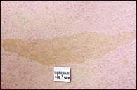
Café au lait (coffee with milk) macules can be congenital, or they may develop in childhood. These flat macules usually occur on the trunk and can have a smooth or irregular border (Figure 2). They range from 0.2 to 4 cm in diameter in infants but can reach 30 cm in diameter in adults.3 The hyperpigmentation is caused by increased melanin in melanocytes and basal keratinocytes.4
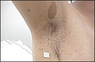
Café au lait macules may be a sign of neurofibromatosis. The diagnosis of this disease requires the presence of at least two of seven criteria established at a National Institutes of Health (NIH) consensus development conference (Table 2).5 The presence of six or more café au lait macules is one of the diagnostic criteria; axillary or groin freckling is another (Figure 3). Hence, the presence of axillary or groin freckling should prompt a search for café au lait macules, and vice versa. If neurofibromatosis is suspected or confirmed, an ophthalmologist can detect optic gliomas and iris hamartomas, which are additional criteria for the diagnosis of neurofibromatosis. The American Academy of Dermatology has practice guidelines based on the NIH definition.5 [Evidence level C, consensus opinion]
Diagnosis requires the presence of two or more of the following:
|
Café au lait macules themselves require treatment only if cosmesis is requested. Surgical or laser treatment by a cosmetic dermatologist may be considered on an individual basis. From a practical standpoint, neurofibromas, which can occur throughout the body, pose more of a cosmetic and functional concern (Figure 4).
Consultation with a geneticist may be prudent because neurofibromatosis is an autosomal dominant condition, although spontaneous mutations cause 50 percent of cases.6 Some patients have café au lait macules without neurofibromatosis; their children are not at increased risk for the disease.
Diffuse Hyperpigmentation
Diffuse hyperpigmentation may have a systemic cause, such as Addison's disease, hyperthyroidism, or hemochromatosis. It also may occur because of a medication side effect.
In Addison's disease, the adrenal glands fail to produce adequate amounts of mineralo-corticoids and glucocorticoids. When elevations of melanocyte-stimulating hormone and adrenocorticotropic hormone (also referred to as corticotropin) levels occur as the pituitary gland tries to stimulate the adrenal glands, melanin production increases, causing hyperpigmentation with a “muddy” appearance.7 The hyperpigmentation is diffuse, but more pronounced in sun-exposed areas and in the perineum, axillae, areolae, palms, and soles.8 Patients with Addison's disease should be treated with mineralocorticoid and glucocorticoid replacement to reduce the drive for excess production of adrenocorticotropic hormone and melanocyte-stimulating hormone.
Hyperthyroidism causes a pattern of hyperpigmentation similar to that in Addison's disease, especially in patients with darker complexions.9 Treatment is specific to the cause of hyperthyroidism and may involve antithyroid medications, surgical resection of the thyroid, or radioactive iodine therapy.
Hemochromatosis, a disorder of iron storage and deposition, can cause a slate-gray hyperpigmentation (because of hemosiderin deposition). Hyperpigmentation is present in 70 percent of affected patients at diagnosis.10 Bronzing resulting from increased melanin production is common, although the mechanism for this effect is not known.11 Hemochromatosis is treated with repeated phlebotomy to reduce excessive iron stores.12
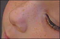
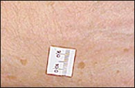
Lesions from Chronic Sun Exposure
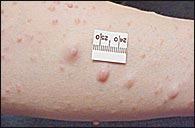
The normal response to solar radiation (or tanning beds) is an increase in melanin production, which causes uniform tanning in most persons but leads to freckling in some. Ephelides, or freckles, are small (usually less than 3 mm in diameter), red or light- to dark-brown macules that appear on sun-exposed areas of the body (Figure 5). Lentigines are tan, brown, or black macules, 2 to 20 mm in diameter, that occur anywhere on the body (Figure 6).
Lentigines have increased numbers of melanocytes that produce dense epidermal deposits of melanin. In contrast to lentigines, ephelides have a normal number of melanocytes but an increased number of melanosomes.13 Because both lentigines and ephelides are benign, differentiation is not critical.
Ephelides and lentigines can be too numerous to count. This factor and their relative uniformity of color help to distinguish them from nevi, which have more serious implications. If lesions are larger than several millimeters or display abnormal coloration, the differential diagnosis includes nevi, which may require surgical removal because of possible malignancy.
Multiple lentigines in the skin, lips, and mucous membranes should raise suspicion for Peutz-Jeghers syndrome. This autosomal dominant syndrome is associated with polyposis of the gastrointestinal tract and an increased risk of cancers of the pancreas, lung, breast, ovary, and uterus.14 [Evidence level B, retrospective cohort study]
Treatment of ephelides and lentigines is for cosmesis only. Avoidance of sun exposure and the use of sunscreens and make-up or moisturizers that contain sunscreens can decrease the development of additional lesions. If desired, patients can be treated with bleaching solutions or with peeling agents by a professional who is experienced in their use, such as a dermatologist. However, results are slow, and complications such as irritation and hyperpigmentation can occur. Bleaching agents include hydroquinone (Eldoquin Forte), which is available in 2 to 4 percent creams and gels. Laser therapy also is an option, but because of the frequency and broad distribution of these lesions, referral to a cosmetic dermatologist is appropriate.
Lesions from Phototoxic Reactions
Hyperpigmentation can be caused by phototoxic reactions from the use of systemic or topical medications (Table 3)15 or from contact with certain plants or foods in conjunction with sun exposure16 (phytophotodermatitis). Initially, patients develop an erythematous allergic reaction. There can be an inflammatory response with lymphocytes, eosinophils, and edema, which can result in a bullous reaction on sun-exposed skin. Over time, hyperkeratosis and melanocytic hyperplasia develop, causing hyperpigmentation.1 Some medications result directly in hyperpigmentation, without sun exposure, in a diffuse pattern17,18 (Table 4).17,19,20 Examples of plant materials and foods that may be associated with phytophotodermatitis include lemons, limes, fig leaves or stems, celery, dill, parsnips, and carrot juice.16
| Anthranilic acids | Miscellaneous | ||
| Meclofenamic acid | Amiodarone (Cordarone) | ||
| Antibiotics | Desipramine (Norpramin) | ||
| Ceftazidime (Fortaz) | Diltiazem (Cardizem) | ||
| Fluoroquinolones | Fibric acid derivatives | ||
| Griseofulvin (Grisactin) | Imipramine (Tofranil) | ||
| Ketoconazole (Nizoral) | Phenothiazines | ||
| Nalidixic acid (NegGram) | Quinidine | ||
| Sulfonamides | Quinine | ||
| Tetracyclines | Sulfite food derivatives | ||
| Trimethoprim (Proloprim; also, in Bactrim, Septra) | Nonsteroidal anti-inflammatory drugs | ||
| Arylpropionic acid derivatives | |||
| Antineoplastic agents | Benoxaprofen (Oraflex) | ||
| Dacarbazine (Dtic-Dome) | Carprofen (Rimadyl) | ||
| Fluorouracil (Adrucil) | Ibuprofen (Motrin) | ||
| Methotrexate | Ketoprofen (Orudis) | ||
| Vinblastine (Velban) | Nabumetone (Relafen) | ||
| Coal tar | Naproxen (Naprosyn) | ||
| Diuretics | Tiaprofenic acid (Surgam) | ||
| Bendroflumethiazide (Naturetin) | Porphyrins | ||
| Furosemide (Lasix) | Psoralens | ||
| Hydrochlorothiazide (Esidrix) | Pyrazolidinediones | ||
| Dyes | Oxyphenbutazone (Phenabid) | ||
| Eosin | Phenylbutazone (Butazolidin) | ||
| Fluorescein | Retinoids | ||
| Methylene blue | Etretinate (Tegison) | ||
| Rose Bengal | Isotretinoin (Accutane) | ||
| Salicylic acids | |||
| Aspirin | |||
| Diflunisal (Dolobid) | |||
| Amiodarone (Cordarone) |
| Amitriptyline (Elavil) |
| Arsenic |
| Bismuth |
| Bleomycin (Blenoxane) |
| Busulfan (Myleran) |
| Clofazimine (Lamprene) |
| Cyclophosphamide (Cytoxan) |
| Daunorubicin (DaunoXome, Cerubidine) |
| Doxorubicin (Adriamycin) |
| Gold (may cause chrysiasis) |
| Mercury |
| Minocycline (Minocin) |
| Nitrogen mustard (topical) |
| Phenothiazines |
| Silver (may cause argyria) |
| Zidovudine (Retrovir) |
Patients with phototoxic reactions should discontinue use of the offending medication or contact with the offending substance. If necessary, exposure to sunlight should be avoided. For cosmetic concerns, phototoxic reactions that fail to resolve spontaneously should be managed similarly to melasma, which is discussed in part II of this article.