Photo Quiz
New-Onset Bullous Rash
Am Fam Physician. 2015 Feb 15;91(4):231-232.
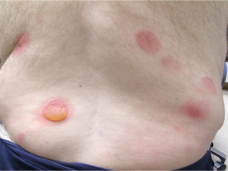
A 63-year-old man presented with an asymptomatic rash that began four days earlier, one day after he was treated for an abscess on his right abdomen. The abscess was treated by incision and drainage, and he was prescribed trimethoprim/sulfamethoxazole because he had a history of methicillin-resistant Staphylococcus aureus infection.
Physical examination revealed multiple large, erythematous, sharply demarcated circular plaques, some with tense bullae in the center, on his trunk and extremities (Figures 1 and 2). There was no mucosal involvement.
Figure 1.

Figure 1.
Figure 2.

Figure 2.
Question
Based on the patient's history and physical examination, which one of the following is the most likely diagnosis?
A. Bullous impetigo.
B. Bullous pemphigoid.
C. Erythema multiforme.
D. Pemphigus vulgaris.
Discussion
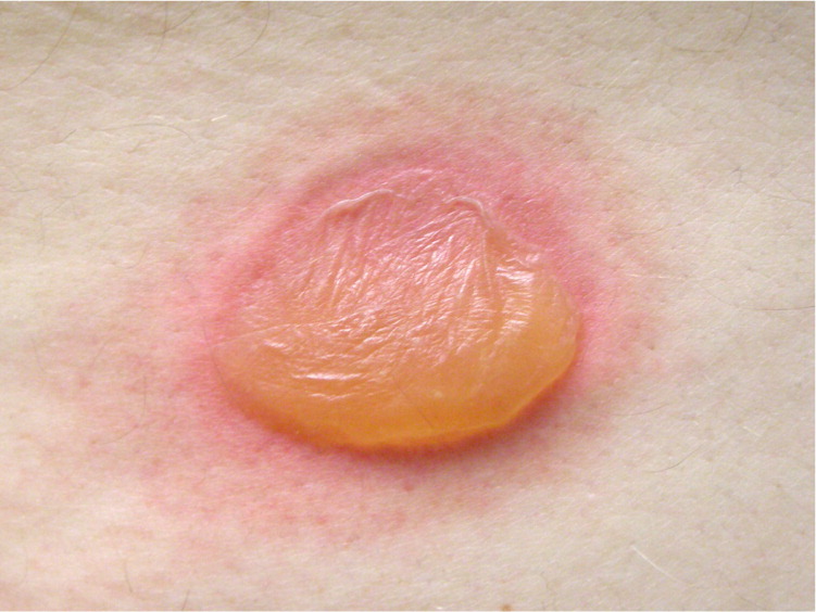
The answer is C: erythema multiforme. Erythema multiforme minus is a self-limited hypersensitivity reaction. It most commonly occurs in response to an antecedent herpes simplex virus infection but may also be triggered by medications, including sulfonamides, barbiturates, penicillins, and phenytoin (Dilantin).1–3 Erythema multiforme majus is a more severe form of the condition also known as Stevens-Johnson syndrome. The diagnosis of erythema multiforme is clinical. Lesions may have a target appearance, and usually begin on the distal extremities and progress proximally.3 Vesicles and bullae sometimes form centrally.3
Erythema multiforme usually causes minimal symptoms and resolves over several weeks without treatment.2 Suspected causal medications should be withdrawn as soon as possible. Oral antihistamines or topical steroids may be used for itching.2 A short course of oral corticosteroids may be helpful in severe cases; however, this is controversial.3 Recurrent cases may be treated with antiviral agents or other immunosuppressant drugs.3
Bullous impetigo is caused by toxin-producing staphylococcal strains and is most common in infants and children.2 Vesicles enlarge rapidly to form bullae containing clear or cloudy fluid. The lesions may expand and rupture, leaving a collarette of moist scale and an erythematous base.2 There is typically little to no surrounding erythema. Diagnosis is usually clinical but may require a culture of fluid from the lesion.4
Bullous pemphigoid is an autoimmune disease characterized by tense bullae on an erythematous, urticarial, or noninflammatory base.5 The bullae are commonly pruritic and may be preceded by a prodromal phase with eczematous, papular, or urticarial skin lesions lasting for weeks.6 Diagnosis is made through biopsy of the lesion and perilesional tissue, with subsequent microscopic evaluation using standard histology and direct immunofluorescent techniques.
Pemphigus vulgaris is an autoimmune disease characterized by flaccid blisters on the skin and mucosal surfaces that rupture easily, leaving painful erosions.7 Diagnosis is made through biopsy of the lesion and perilesional tissue.
Summary Table
Condition | Characteristics |
---|---|
Bullous impetigo | Rapid onset of bullae containing clear or cloudy fluid, most common in infants and children; ruptured lesions may leave a collarette of moist scale and an erythematous base |
Bullous pemphigoid | Tense bullae on an erythematous, urticarial, or noninflammatory base; commonly pruritic and may be preceded by a prodromal phase with eczematous, papular, or urticarial skin lesions |
Erythema multiforme | Hypersensitivity reaction often to an infection or a medication; asymptomatic rash consisting of target-like lesions that commonly begin on the distal extremities and progress proximally |
Pemphigus vulgaris | Autoimmune disease characterized by flaccid blisters on skin or mucosal surfaces; lesions may rupture easily, leaving painful erosions |
REFERENCES
show all references1. Odom RB, James WD, Berger TG, eds. Andrews' Diseases of the Skin: Clinical Dermatology. 9th ed. Philadelphia, Pa.: W.B. Saunders Co.; 2000....
2. Habif TP, ed. Clinical Dermatology: A Color Guide to Diagnosis and Therapy. 5th ed. Edinburgh, Scotland: Mosby; 2010.
3. Lamoreux MR, Sternbach MR, Hsu WT. Erythema multiforme. Am Fam Physician. 2006;74(11):1883–1888.
4. Baddour LM. Impetigo. UpToDate. http://www.uptodate.com/contents/impetigo (subscription required). Accessed August 16, 2013.
5. DiZenzo G, Marazza G, Borradori L. Bullous pemphigoid: physiopathology, clinical features and management. Adv Dermatol. 2007;23:257–288.
6. Kasperkiewicz M, Zillikens D, Schmidt E. Pemphigoid diseases: pathogenesis, diagnosis, and treatment. Autoimmunity. 2012;45(1):55–70.
7. Hertl M, Sitaru C. UpToDate. http://www.uptodate.com/contents/pathogenesis-clinical-manifestations-and-diagnosis-of-pemphigus (subscription required). Accessed August 16, 2013.
This series is coordinated by John E. Delzell, Jr., MD, MSPH, Assistant Medical Editor.
A collection of Photo Quiz published in AFP is available at https://www.aafp.org/afp/photoquiz.
Previously published Photo Quizzes are now featured in a mobile app. Get more information at https://www.aafp.org/afp/apps.
The editors of AFP welcome submissions for Photo Quiz. Guidelines for preparing and submitting a Photo Quiz manuscript can be found in the Authors' Guide at https://www.aafp.org/afp/photoquizinfo. To be considered for publication, submissions must meet these guidelines. E-mail submissions to afpphoto@aafp.org.
Copyright © 2015 by the American Academy of Family Physicians.
This content is owned by the AAFP. A person viewing it online may make one printout of the material and may use that printout only for his or her personal, non-commercial reference. This material may not otherwise be downloaded, copied, printed, stored, transmitted or reproduced in any medium, whether now known or later invented, except as authorized in writing by the AAFP. Contact
afpserv@aafp.org for copyright questions and/or permission requests.
Want to use this article elsewhere? Get Permissions
More in AFP
Editor's Collections
Related Content
More in Pubmed
MOST RECENT ISSUE
Email Alerts
Don't miss a single issue. Sign up for the free AFP email table of contents.