
Am Fam Physician. 2015;91(11):791-792
Author disclosure: No relevant financial affiliations.
A 59-year-old man presented with fever, chills, and a pruritic rash that had generalized peeling and underlying redness. The rash began one month prior as small papules on his arms and then spread across his entire body, including his palms and soles. He also had pain in his hands. The rash did not improve after treatment with a topical medium-potency steroid, but subsequently responded to a course of oral prednisone. His medical history was significant for hypertension and hyperlipidemia. He was taking hydrochlorothiazide and rosuvastatin (Crestor). He did not have any sick contacts and was not using any new soap, lotion, or detergent.
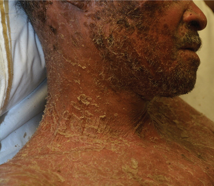
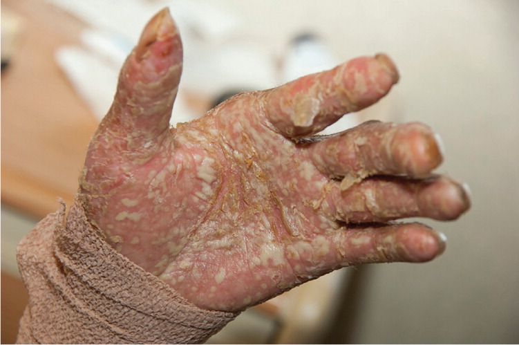
On physical examination, he was ill appearing and febrile. His heart rate was 120 beats per minute, and his blood pressure was 90/50 mm Hg. The skin rash (Figure 1) consisted of diffuse pustules on an erythematous background and involved more than 85% of his body surface area, including his head, trunk, and upper and lower extremities. There was a thick scale, including on the palms and soles (Figure 2). Bacterial and viral culture results were negative.
Question
Discussion
The answer is C: erythrodermic psoriasis. Characteristic features include a diffuse, bright-red rash and desquamation with superficial scaling and often exudation. This is unlike plaque psoriasis, a common variant of psoriasis, which has thick, adherent white scales. The average age of onset is 50 years.1 The erythrodermic form is considered the most active psoriatic process with increased proliferation. This results in a loss of keratinocyte maturation and abnormal keratinocytes that are loosely bound and more easily shed. In this patient, a skin biopsy showed dermal lymphohistiocytic inflammation with occasional eosinophils.
Patients with erythrodermic psoriasis can have excessive heat loss from generalized vasodilation of cutaneous vessels, leading to shivering and hypothermia. Vasodilation can lead to loss of protein and extremity edema.2 Patients with this condition also have signs of systemic illness, including generalized pruritus, fever, chills, dehydration, and lymphadenopathy.1 Erythrodermic psoriasis usually arises from pustular psoriasis but can be triggered by sunburn, human immunodeficiency virus infection, systemic infections, malignancies (mainly lymphomas), and even chronic plaque psoriasis.3 It can also arise after the withdrawal of systemic steroids.1,3 Treatment occurs in the intensive care unit and includes fluid and electrolyte management, and treatment for sepsis.2
Acute generalized exanthematous pustulosis is a rare, drug-induced eruption that is characterized by the sudden appearance of dozens of sterile, nonfollicular, small pustules on erythematous and edematous skin.4 It most commonly occurs with the use of antibiotics (penicillins and macrolides), calcium channel blockers, and antimalarials. Fever and neutrophilia are present, and sometimes there is mild nonerosive mucosal involvement. One-third of patients have peripheral eosinophilia.4 Within a few days of stopping the causative drug, pustules spontaneously disappear. The desquamation and reaction fully resolve within 15 days.4
Allergic contact dermatitis manifests as erythematous vesicles and bullae with scaling and relatively well-demarcated borders on exposed skin, typically along the hands, face, and neck, although any area can be affected. Pruritus is the most common symptom.5
Pityriasis rubra pilaris is an inflammatory condition of uncertain etiology. The rash is typically erythematous with salmon-colored plaques. It is clinically distinguished from plaque psoriasis by the presence of red-orange, palmoplantar, keratotic follicular papules and islands of well-demarcated sparing on the trunk.6 Autoimmune diseases, infections, or malignancies are possible triggers.
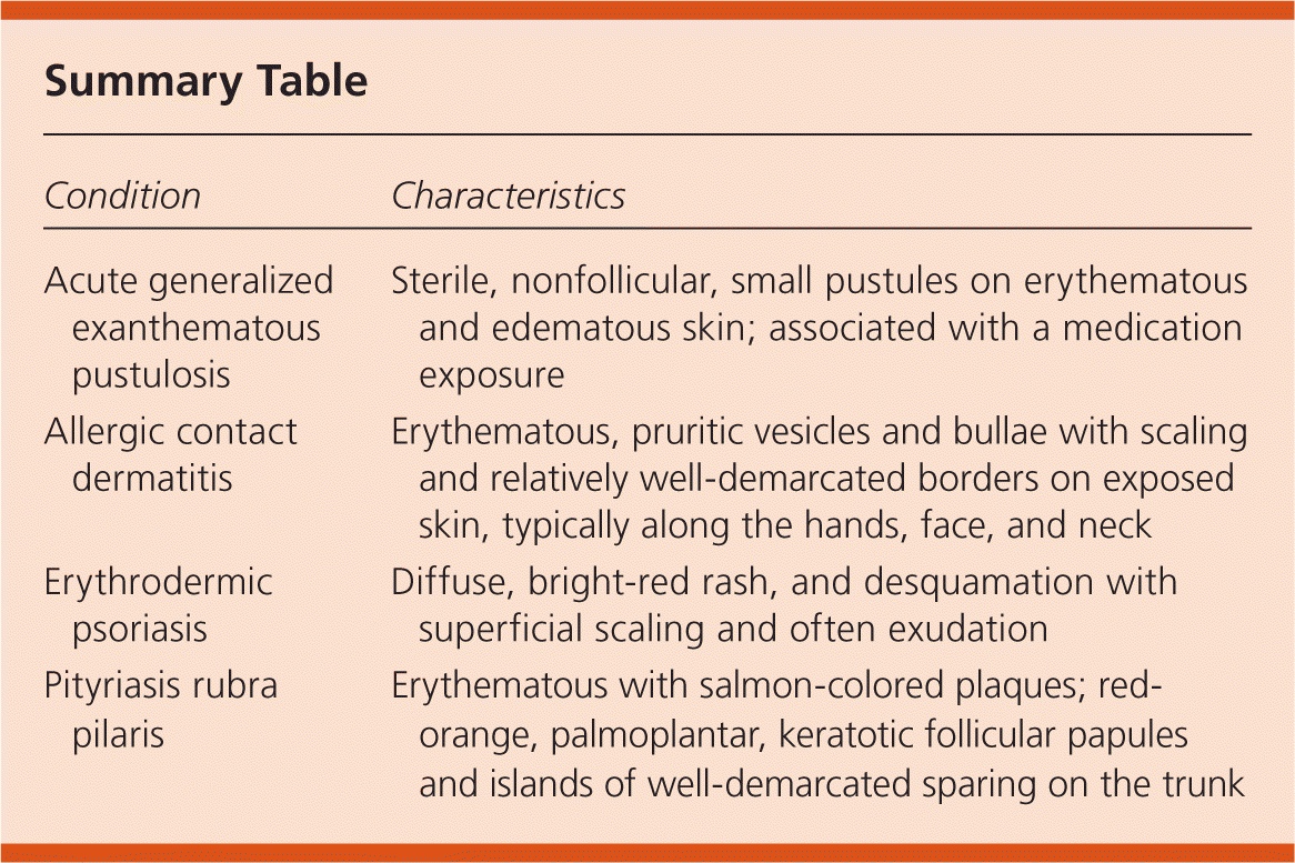
| Condition | Characteristics |
|---|---|
| Acute generalized exanthematous pustulosis | Sterile, nonfollicular, small pustules on erythematous and edematous skin; associated with a medication exposure |
| Allergic contact dermatitis | Erythematous, pruritic vesicles and bullae with scaling and relatively well-demarcated borders on exposed skin, typically along the hands, face, and neck |
| Erythrodermic psoriasis | Diffuse, bright-red rash, and desquamation with superficial scaling and often exudation |
| Pityriasis rubra pilaris | Erythematous with salmon-colored plaques; red-orange, palmoplantar, keratotic follicular papules and islands of well-demarcated sparing on the trunk |
