
Am Fam Physician. 2015;92(5):387-388
Author disclosure: No relevant financial affiliations.
A man presented with a rash that appeared one to two days after he used a hot tub. He developed chills and a fever of 102°F (39°C) the day after the rash appeared. The rash continued to spread and the fever persisted even though he had been taking oral trimethoprim/sulfamethoxazole for two days.
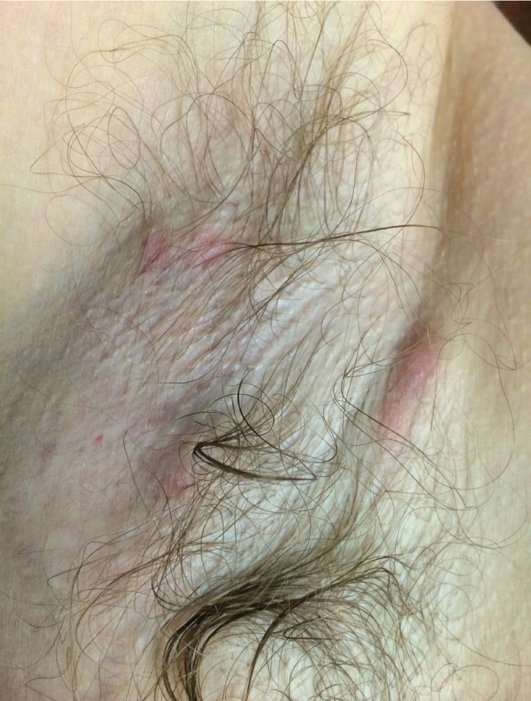
Physical examination revealed four or five superficial, tender, isolated, painful, erythematous nodules in bilateral axilla that were fluctuant and 0.5 to 1 cm in size (Figure 1). Incision and drainage produced a small amount of thick, purulent exudate from each nodule. The erythematous nodules on his abdomen were 1 to 3 mm in size and were not fluctuant. There was a pustule with surrounding erythema on his right calf.
Question
Discussion
The answer is B: P. aeruginosa. The recent use of a hot tub preceding a painful superficial rash is the key feature suggesting P. aeruginosa infection. P. aeruginosa can live freely in the environment and is found in natural water sources, such as lakes and rivers. It rarely affects drinking water but may colonize in plumbing fixtures on biofilms. P. aeruginosa is often found in hot tubs, likely because of the higher temperature and aeration of the water.1
Infection with P. aeruginosa, a gram-negative bacterium, is uncommon in immunocompetent patients. When infection does occur, the severity can vary. Minor skin infections include a maculopapular erythematous rash manifesting as cellulitis or cutaneous nodules. Severe infections can result in tissue necrosis. There may be a few to many lesions, usually located on the axilla, groin, or trunk.2
P. aeruginosa is periodically identified as the pathogen in otitis externa and dermatitis outbreaks that are associated with pool or hot tub use. Insufficient chlorine use and pH monitoring are usually the cause of these outbreaks.3
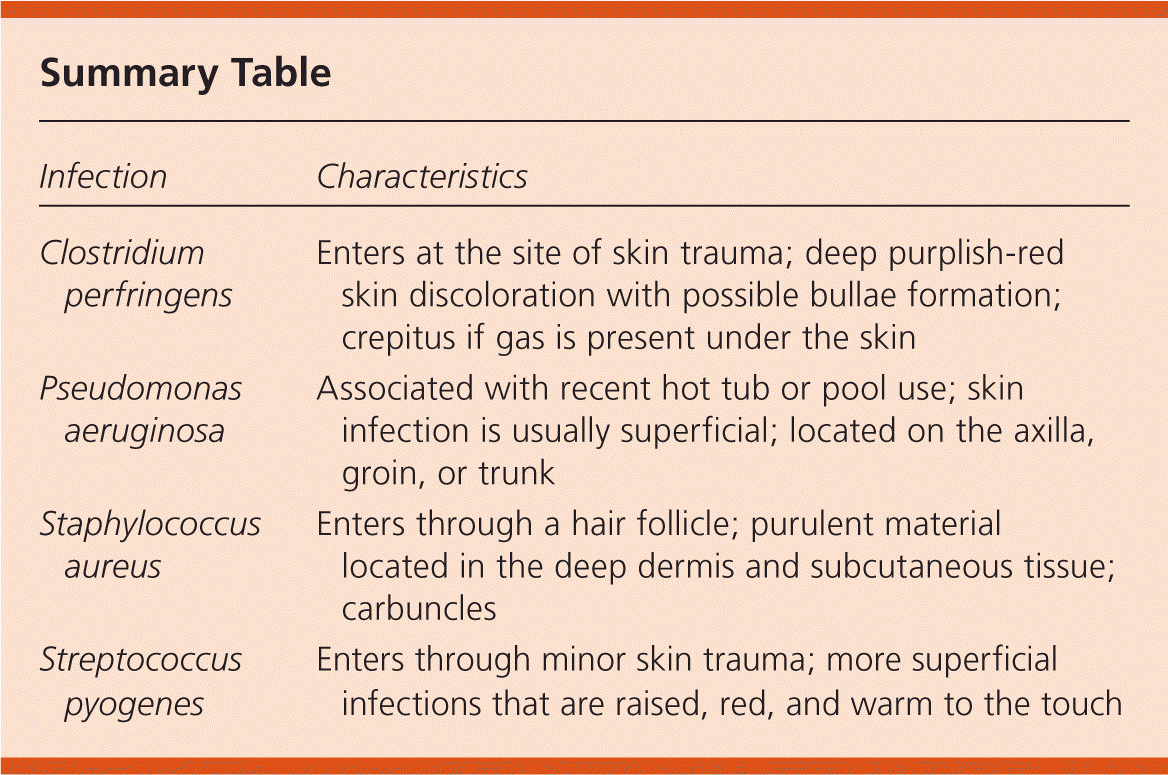
C. perfringens infection can cause myonecrosis, or gas gangrene. The infection is typically associated with skin trauma. The skin appears deep purplish-red and taut, and is very tender. Hemorrhagic bullae can develop. Often, gas can be palpated in the skin as crepitus. Systemic signs of infection develop rapidly, often within 24 hours.2
S. aureus can cause purulent material to collect in the deep dermis and subcutaneous tissue. A hair follicle is typically the entry point. When several areas of infection join, they form a carbuncle. These tracts of infection are palpable. This infection often leads to repeated development of abscesses. About 20% to 40% of the population is colonized with S. aureus, and it is unknown why only some of these individuals develop repeated infections.2
S. pyogenes typically forms a diffuse spreading superficial skin infection. It is usually associated with mild trauma that is often unrecognized by the patient. The infection develops rapidly with spreading edema, erythema, and heat. When the infected skin is raised with a noted border of demarcation, it is known as erysipelas. S. pyogenes infection is more common in older persons and children.2
| Infection | Characteristics |
|---|---|
| Clostridium perfringens | Enters at the site of skin trauma; deep purplish-red skin discoloration with possible bullae formation; crepitus if gas is present under the skin |
| Pseudomonas aeruginosa | Associated with recent hot tub or pool use; skin infection is usually superficial; located on the axilla, groin, or trunk |
| Staphylococcus aureus | Enters through a hair follicle; purulent material located in the deep dermis and subcutaneous tissue; carbuncles |
| Streptococcus pyogenes | Enters through minor skin trauma; more superficial infections that are raised, red, and warm to the touch |
