A 77-year-old woman presented with a longstanding history of daily pain and stiffness in multiple joints. She reported morning stiffness in her shoulders, knees, dorsal feet, and metacarpophalangeal joints that improved with hot showers. She did not have acute attacks of pain or constitutional symptoms such as fever, chills, or weight changes.
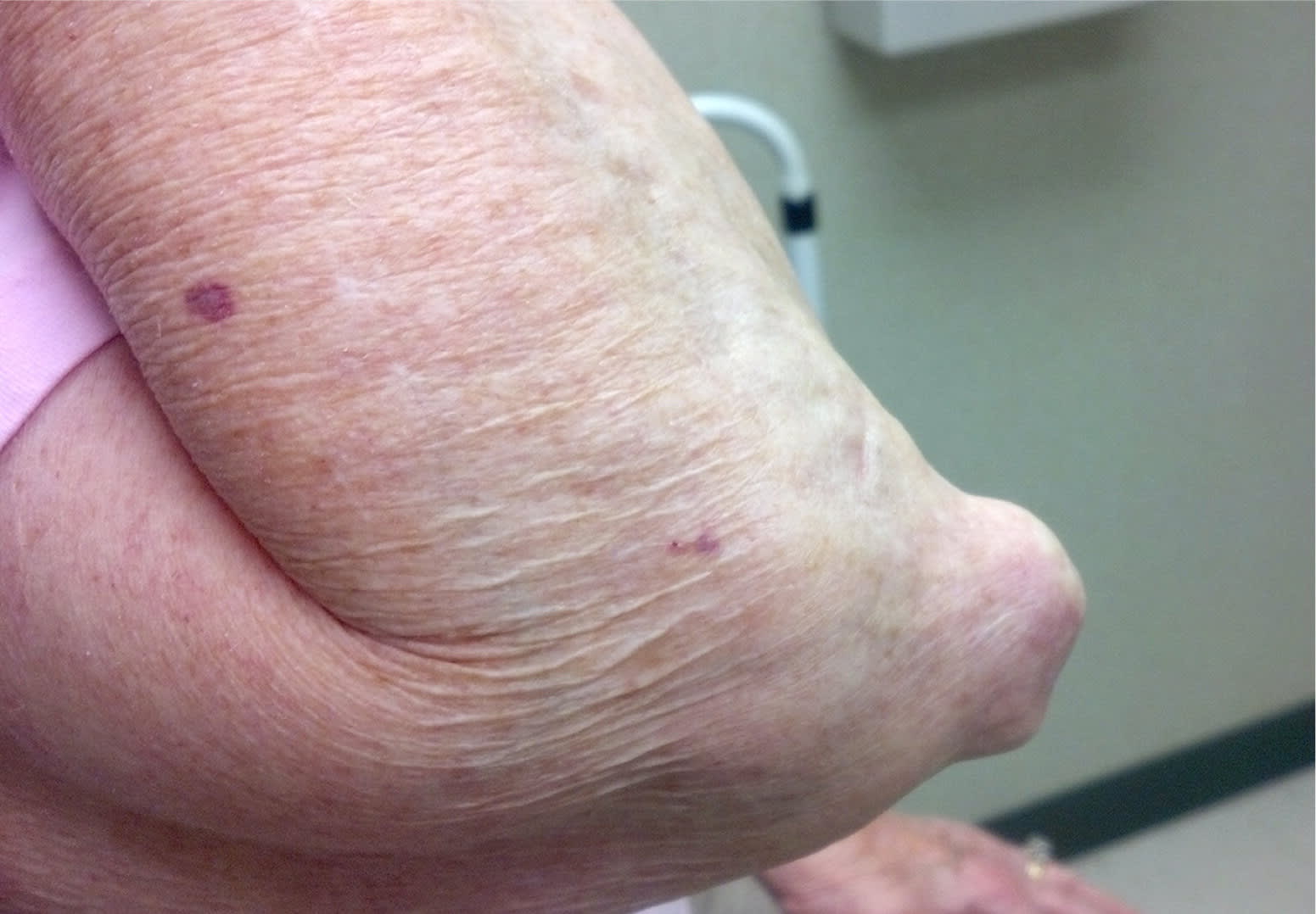
On physical examination, she had symmetric swelling, warmth, and tenderness of the second, third, and fourth metacarpophalangeal joints. She also had bilateral irregularly shaped, nontender, firm, fixed nodules over the extensor surfaces of her elbows (Figure 1).
Figure 1.
Question
Based on the patient's history and physical examination findings, which one of the following is the most likely diagnosis?
A. Epidermoid cysts.
B. Olecranon bursitis.
C. Rheumatoid nodules.
D. Tophaceous gout.
Discussion
The answer is C: rheumatoid nodules. Rheumatoid arthritis is a symmetric polyarthritis that typically involves the small joints of the hands and feet, although any joint with a synovial lining may be affected. Classically, it begins with the gradual onset of pain and stiffness that is worse in the morning. The joints are usually affected for an hour or more, and there is improvement with activity.1 The nodules are usually found on areas exposed to chronic pressure but may occur within other organ systems, including the lung, heart, and rarely the central nervous system.
Rheumatoid nodules are the most common cutaneous finding in rheumatoid arthritis, occurring in 30% to 40% of patients with the disease.2 The exact etiology is unknown. They are composed of inflammatory cells, fibrin, and necrotic proteinaceous debris.3 Most patients with rheumatoid nodules are seropositive for rheumatoid factor and anticyclic citrullinated peptide antibodies.4 Patients with rheumatoid nodules typically have a more severe course and more rapid progression of rheumatoid arthritis than patients without nodules.5 Nodules range from 2 mm to 5 cm and are firm, usually nontender, and mobile, although they may become fixed to the underlying periosteum.6 Occasionally, nodules may be painful or large enough to limit range of motion or compress nearby nerves, necessitating treatment. Treatments include glucocorticoid injections,7 and occasionally surgical excision for recalcitrant cases.8
Epidermoid cysts are benign, mobile dermal growths that often contain a central punctum. They are filled with keratinaceous debris. Intact cysts may be removed by simple excision, whereas inflamed cysts may be injected with corticosteroids and excised when asymptomatic.9
Olecranon bursitis is characterized by the fluctuant collection of fluid in the olecranon bursa due to trauma, chronic pressure, overuse, or other rheumatic conditions. Signs of inflammation and tenderness depend on the acuity of the etiologic process. Unless a secondary infection is suspected, treatment consists of joint protection and compression with or without aspiration.
Tophaceous gout is considered the end-stage of monosodium urate crystal deposition. Tophi are firm, nontender nodules often with associated chronic inflammation and destructive changes in nearby tissues. Although the tophi are usually not painful, gout is typically characterized by the onset of discrete attacks of monoarticular arthritis interspersed with asymptomatic periods.
Summary Table
| Condition | Characteristics |
|---|---|
| Epidermoid cyst | Benign, mobile dermal growths that often contain a central punctum; filled with keratinaceous debris |
| Olecranon bursitis | Fluctuant collection of fluid in the olecranon bursa due to trauma, chronic pressure, overuse, or other rheumatic conditions |
| Rheumatoid nodule | Firm, nontender, mobile or fixed nodule that develops over pressure points as an extra-articular manifestation of rheumatoid arthritis |
| Tophaceous gout | Firm, nontender nodule caused by the repeated deposition of monosodium urate crystals in soft tissues |
