
Am Fam Physician. 2017;96(11):739-741
Author disclosure: No relevant financial affiliations.
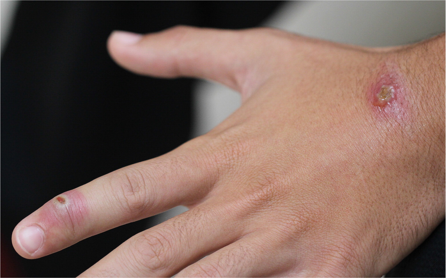
A healthy 17-year-old presented with a one-week history of slowly progressing, painless skin lesions on the dorsum of the hand and the index finger. Approximately three weeks before the lesions appeared, he received a minor laceration on his hand while skinning slaughtered sheep at a farm. No topical medications had been applied to the lesions. The patient had no constitutional symptoms or other lesions.
Physical examination revealed an 8-mm, weeping nodule with surrounding induration and erythema on the left dorsal hand. A lesion on the index finger had an erythematous center with circumscribed hypopigmentation within a surrounding area of erythema (Figure 1).
Question
Based on the patient's history and physical examination findings, which one of the following is the most likely diagnosis?
A. Contagious ecthyma (orf).
B. Cutaneous anthrax.
C. Mycobacterium marinum infection.
D. Nocardia infection.
E. Sporothrix schenckii infection.
Discussion
The answer is A: contagious ecthyma (orf), which is caused by a parapoxvirus. It is primarily transmitted from infected sheep or goats through direct contact or contaminated fomites. Orf is typically an occupational skin disease, but household exposures can occur with animal slaughtering or religious animal sacrifices. There have also been cases of children contracting orf after visiting petting zoos and livestock fairs.1,2 Hands are the most common site of infection and clinical presentation.
In immunocompetent patients, there are six distinct stages of orf 2,3: (1) three to seven days after inoculation, a painless erythematous macule appears and progresses into a papule (maculopapular stage); (2) the lesion develops a red center with a surrounding white ring and a red halo (targetoid stage), as depicted in Figure 1; (3) a weeping nodule forms (acute or weeping nodule stage); (4) the lesion begins to dry and develop a thin yellow crust and small black dots (regenerative stage); (5) the lesion becomes papilloma-like (papillomatous stage); and (6) the lesion crusts over and resolves (regressive stage), usually without scarring.
Diagnosis of orf is usually clinical; however, histologic examination with or without electron microscopy or polymerase chain reaction testing can help differentiate it from other skin infections.5 Orf resolves spontaneously in four to eight weeks and requires only supportive care to prevent secondary infection. Topical imiquimod (Aldara) can be used to shorten the duration of the disease, particularly in patients who are immunocompromised, who can have more pronounced and extended disease courses.6
Bacillus anthracis, which causes anthrax, is most often transmitted via direct contact with hides or wools of infected sheep, cows, horses, or goats (woolsorter's disease). Cutaneous anthrax presents as a purpuric macule or papule that ulcerates and forms a painless, black eschar with surrounding erythema. Systemic symptoms occasionally occur. Mortality from cutaneous anthrax can be as high as 20% if not treated appropriately.4
Humans can be infected with Mycobacterium marinum, a free-living, nontuberculous mycobacterium, through exposure to contaminated fresh or salt water, or fish carcasses. It is an occupational disease among fish handlers. After an incubation period of two to six weeks, a painless red papule slowly progresses into violaceous, nodular plaques that can be pustular and ulcerated.7 Systemic symptoms are not usually present.4
Nocardia species are a group of filamentous, gram-positive, acid-fast organisms that are found ubiquitously in the soil. They are opportunistic pathogens in persons who are immunocompromised, but cutaneous nocardiosis can also occur in immunocompetent patients.4 The infection may present in three forms: a painless, enlarging nodule with purulent discharge; a crusted papule or abscess; or superficial cellulitis.8
Sporothrix schenckii is a dimorphous fungus that causes sporotrichosis (rose gardener's disease). Infection occurs from trauma and exposure to contaminated sources, such as soil or plants.9 Cutaneous sporotrichosis presents as a papule at the site of injury. The lesion can become nodular or ulcerate with associated erythema and may have purulent drainage. Additional lesions may present along the path of lymphatic drainage (sporotrichoid pattern). The infection causes mild pain without systemic symptoms.4
| Condition | Characteristics |
|---|---|
| Contagious ecthyma (orf) | Painless maculopapular lesions progressing through six stages; systemic symptoms are uncommon |
| Cutaneous anthrax | Purpuric macule or papule that ulcerates and forms a painless, black eschar with surrounding erythema; occasionally accompanied by systemic symptoms |
| Mycobacterium marinum infection | Painless red papule slowly progressing to violaceous, nodular plaques that can be pustular and ulcerated |
| Nocardia infection | Painless, enlarging nodule with purulent discharge, crusted papule or abscess, or superficial cellulitis |
| Sporothrix schenckii infection | Papule that can become nodular and ulcerate with associated erythema; lesions may present along the path of lymphatic drainage; mild pain without systemic symptoms |
The contents of this publication are the sole responsibility of the authors and do not necessarily reflect the views, assertions, opinions, or policies of the Uniformed Services University of the Health Sciences, the Department of Defense, or the Departments of the Army, Navy, or Air Force. Mention of trade names, commercial products, or organizations does not imply endorsement by the U.S. government.
