A 44-year-old man presented with worsening scrotal swelling that began two weeks prior. He also had sharp, constant pain that radiated to both legs (left more than right) and his buttocks. He rated it as 10 out of 10 on a pain scale. The pain worsened with urination and was minimally relieved by over-the-counter ibuprofen. He had never had a similar episode and had no injuries. His sexual history was significant for multiple male partners since 21 years of age. He had untreated HIV infection, which was diagnosed in 2007. He had no other significant medical history.
Physical examination revealed diffuse scrotal edema and tenderness and multiple black blisters on the scrotum and perianal area (Figure 1). The lesions were warm to the touch and erythematous. He had no penile tenderness or discharge. His white blood cell count was 16.82 per μL (0.02 × 10⁹ per L) with an absolute CD4 cell count of 506 per μL (0.50 × 10⁹ per L).
FIGURE 1

Question
Based on the patient's history and physical examination findings, which one of the following is the most likely diagnosis?
A. Gas gangrene.
B. Kaposi sarcoma.
C. Necrotizing fasciitis.
D. Pyoderma gangrenosum.
E. Pyomyositis.
Discussion
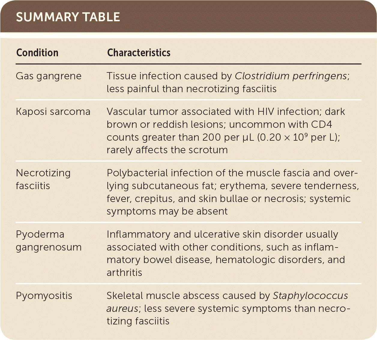
The answer is C: necrotizing fasciitis, known as Fournier gangrene when affecting the perineum or genital regions. Fournier gangrene is a rare life-threatening necrotizing fasciitis caused by a polybacterial infection of the muscle fascia and overlying subcutaneous fat, which has a relatively poor blood supply. Necrotizing fasciitis usually presents as erythema, severe tenderness, fever, crepitus, and skin bullae or necrosis. However, systemic symptoms such as fever may be absent, as in this patient.1 Necrotizing fasciitis is an emergency requiring surgical debridement. Broad-spectrum antibiotics should be started empirically. Diagnosis is established by surgical exploration and tissue biopsy. The mortality rate is high (20% to 88%) even with treatment, and the condition can progress rapidly if not treated appropriately.2
Gas gangrene is a tissue infection caused by Clostridium perfringens, a gram-positive rod bacterium. Although necrotizing fasciitis and gangrene both may lead to gas formation in the tissues, gas gangrene is more common following traumatic injuries. Gas gangrene usually causes less pain than necrotizing fasciitis.3 Treatment of gas gangrene may require amputation.
Kaposi sarcoma is a vascular tumor associated with HIV infection. The skin lesions are dark brown or reddish and may look similar to the bullae of necrotizing fasciitis. Kaposi sarcoma does not usually occur with CD4 counts greater than 200 per μL (0.20 × 10⁹ per L). Lesions may occur on multiple areas, but scrotal lesions are rare.4
Pyoderma gangrenosum is an inflammatory and ulcerative skin disorder. It may be challenging to distinguish from necrotizing fasciitis because it causes similar skin lesions, which quickly progress to ulcers. Most cases are associated with other diseases, such as inflammatory bowel disease, hematologic disorders, and arthritis.5
Pyomyositis is a skeletal muscle abscess caused by Staphylococcus aureus. Magnetic resonance imaging can distinguish it from necrotizing fasciitis because of the type of tissue involved (muscle as opposed to fascia and adipose tissue). Pyomyositis has less severe systemic symptoms than necrotizing fasciitis and does not require emergent surgery.6
SUMMARY TABLE
| Condition | Characteristics |
|---|---|
| Gas gangrene | Tissue infection caused by Clostridium perfringens; less painful than necrotizing fasciitis |
| Kaposi sarcoma | Vascular tumor associated with HIV infection; dark brown or reddish lesions; uncommon with CD4 counts greater than 200 per μL (0.20 × 10⁹ per L); rarely affects the scrotum |
| Necrotizing fasciitis | Polybacterial infection of the muscle fascia and overlying subcutaneous fat; erythema, severe tenderness, fever, crepitus, and skin bullae or necrosis; systemic symptoms may be absent |
| Pyoderma gangrenosum | Inflammatory and ulcerative skin disorder usually associated with other conditions, such as inflammatory bowel disease, hematologic disorders, and arthritis |
| Pyomyositis | Skeletal muscle abscess caused by Staphylococcus aureus; less severe systemic symptoms than necrotizing fasciitis |
