
Am Fam Physician. 2020;101(4):241-242
Author disclosure: No relevant financial affiliations.
A 63-year-old man presented for a wellness visit where he reported worsening dysphagia for solids for the past two years. He also had sore throat, dyspnea, cough, heartburn, and wheezing. He did not have weight loss, hematemesis, melena, or chest pain. His medical history was significant for gastroesophageal reflux disease that responded to over-the-counter antacids.
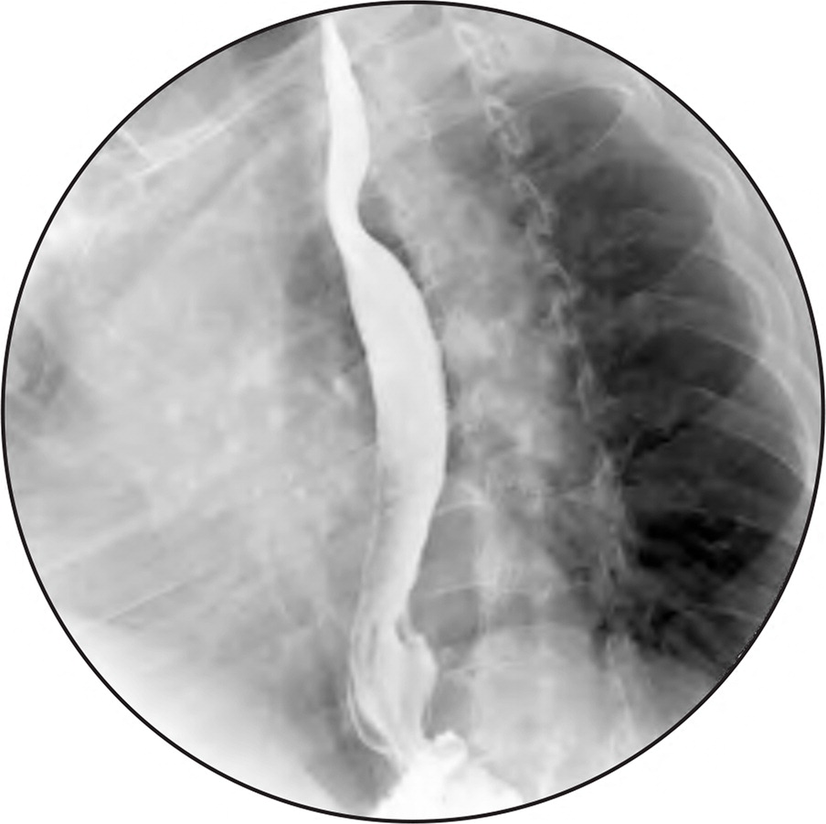
Physical examination findings were normal. A barium study was performed (Figure 1).
Question
Discussion
The answer is B: congenital anomaly of the aortic arch. The barium esophagram shows a filling defect in the esophagus that was noted at the level of the aortic arch, suggesting a possible aberrant vessel of the aorta. Computed tomography (CT) confirmed an aberrant right subclavian artery that passed posterior to the esophagus. An aberrant right subclavian artery, or arteria lusoria, is present in 0.5% to 2.5% of the population.1 Dysphagia is the most common symptom, although only 10% of patients with the anomaly are symptomatic.2 The mean age of presentation is 50 years.3 Possible explanations for this patient’s later presentation include aging-associated stiffening of the esophagus and atherosclerosis of the vessel causing compression of surrounding structures.4
The patient was referred for vascular surgery. He underwent right carotid subclavian transposition followed by ligation of the aberrant right subclavian artery.
An aortic arch aneurysm may compress the esophagus, causing dysphagia. Pain is the most common presenting symptom. CT will confirm dilatation of the aortic arch.
A diffuse esophageal spasm is a common cause of dysphagia for solids and liquids. It usually has a characteristic corkscrew motility pattern on a barium esophagram.
Esophageal webs may cause dysphagia for solids. They may be visualized on a barium esophagram, usually as a thin projection into the lumen.
Mediastinal masses usually appear on a barium esophagram as anterior protrusions into the esophageal lumen between the esophagus and airway. This patient’s esophagram showed a posterior rather than anterior protrusion.
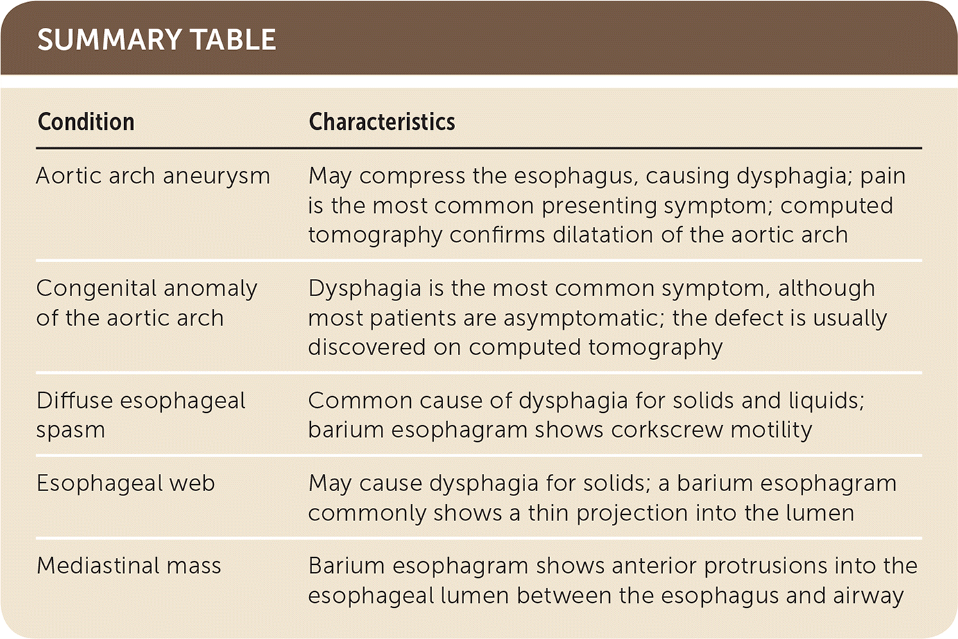
| Condition | Characteristics |
|---|---|
| Aortic arch aneurysm | May compress the esophagus, causing dysphagia; pain is the most common presenting symptom; computed tomography confirms dilatation of the aortic arch |
| Congenital anomaly of the aortic arch | Dysphagia is the most common symptom, although most patients are asymptomatic; the defect is usually discovered on computed tomography |
| Diffuse esophageal spasm | Common cause of dysphagia for solids and liquids; barium esophagram shows corkscrew motility |
| Esophageal web | May cause dysphagia for solids; a barium esophagram commonly shows a thin projection into the lumen |
| Mediastinal mass | Barium esophagram shows anterior protrusions into the esophageal lumen between the esophagus and airway |
