
Am Fam Physician. 2020;101(6):375-376
Author disclosure: No relevant financial affiliations.
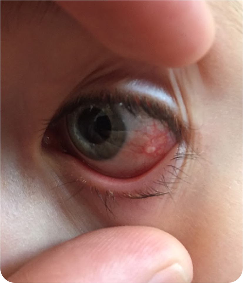
A 10-year-old girl presented with a lesion on her eye that appeared one month prior. The lesion was not painful, and she had no changes in her vision. Her medical history was unremarkable, including no eye trauma or injury. She had no history of contact lens use.
Physical examination revealed a smooth, clear, fluid-filled lesion on the lateral aspect of the left eye (Figure 1). The surrounding conjunctiva was erythematous. The physical examination was otherwise normal.
Question
Discussion
The answer is B: conjunctival cyst. Conjunctival cysts are thin-walled inclusion cysts that progress slowly.1 They commonly occur in the lower fornix at the junction of the conjunctival membranes lining the inside of the eyelid and the eyeball.2 The cysts are lined with epithelial cells and often filled with clear fluid, although they may appear to be solid on visualization.3
Conjunctival cysts are typically asymptomatic and may resolve spontaneously, although the probability of spontaneous resolution is unknown. As the cysts grow, they can become symptomatic and may cause disfigurement, discomfort or foreign body sensation, dry eyes, reduced extraocular motility, or difficulty with eye closure.1 Primary cysts are congenital and often remain hidden in the fornix until they increase in size and become symptomatic.1 Secondary cysts are due to trauma, degeneration, or rarely parasitic infections such as cysticercosis.1
Asymptomatic conjunctival cysts do not need treatment and can be monitored for spontaneous resolution. Supportive treatments, such as lubricating eye drops or ointments, can be used for mild symptoms. Patients should be referred to an ophthalmologist when supportive treatment does not adequately control symptoms.1,3 The cysts should not be ruptured in the clinic.
A conjunctival bleb occurs when there is a direct connection of the anterior chamber to the subconjunctival space due to trauma. This is often associated with glaucoma surgery. It presents as a white, rounded growth often on the superior aspect of the conjunctiva under the upper eyelid. These lesions should not be punctured or deflated because this could lead to hypotonia of the eye or endophthalmitis (inflammation of the tissues or fluids inside the eye).3
A pinguecula is a localized, elevated, yellow-white area on the bulbar conjunctiva near the nasal limbus (border of the cornea and sclera). This is a common condition that is typically asymptomatic and does not require treatment. If the diagnosis is uncertain, the patient should be referred to rule out conjunctival neoplasia.
A pterygium presents as a triangular-shaped growth of the conjunctival and fibrovascular tissue over the limbus and the superficial cornea. Symptoms of irritation can be treated with lubricant eye drops or a mild steroid. In more severe cases, visual acuity can be affected by the onset of an astigmatism or invasion of the pupil. Patients with vision changes or cosmetic concerns should be referred to an ophthalmologist.3
A pyogenic granuloma presents as a fleshy, fast-growing, red, pedunculated vascular mass. It is an inflammatory lesion of the conjunctiva and often appears after surgery or minor trauma to the conjunctiva. Steroid eye drops can clear the granuloma, but excision and cauterization may be needed.3
| Condition | Characteristics |
|---|---|
| Conjunctival bleb | Direct connection of the anterior chamber to the subconjunctival space due to trauma or glaucoma surgery; appears as a white, rounded growth often on the superior aspect of the conjunctiva under the upper eyelid |
| Conjunctival cyst | Thin-walled inclusion cyst filled with clear fluid often located in the lower fornix; may be asymptomatic |
| Pinguecula | Localized, elevated, yellow-white area on the bulbar conjunctiva |
| Pterygium | Triangular-shaped growth of the conjunctival and fibrovascular tissue over the limbus and superficial cornea |
| Pyogenic granuloma | Fleshy, fast-growing, red, pedunculated vascular mass that often appears after surgery or minor trauma to the conjunctiva |
The opinions and assertions contained herein are those of the authors and are not to be construed as official or as reflecting the views of the Uniformed Services University of the Health Sciences, U.S. Air Force Medical Department, the Air Force at large, or the Department of Defense.
