
Am Fam Physician. 2020;102(8):497-498
Author disclosure: No relevant financial affiliations.
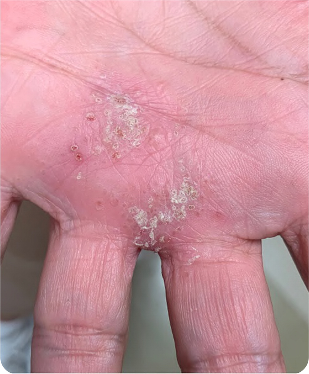
A 61-year-old patient presented with a pruritic rash on the palm of the hand. The patient first noticed the rash one month earlier after working in the garden and described the initial rash as scratches and bumps. The pruritus progressively worsened, and the rash did not improve with hydrocortisone ointment.
Physical examination revealed crusted papular lesions on a mildly erythematous base located on the palm of the left hand (Figure 1). The lesion was approximately 2 × 2 cm in size. No lymphadenopathy was present. The patient had no joint swelling or tenderness. Examination findings were otherwise normal.
Question
Based on the patient's history and physical examination findings, which one of the following is the most likely diagnosis?
A. Actinomycosis.
B. Cellulitis.
C. Cutaneous anthrax.
D. Cutaneous nocardiosis.
E. Sporotrichosis.
Discussion
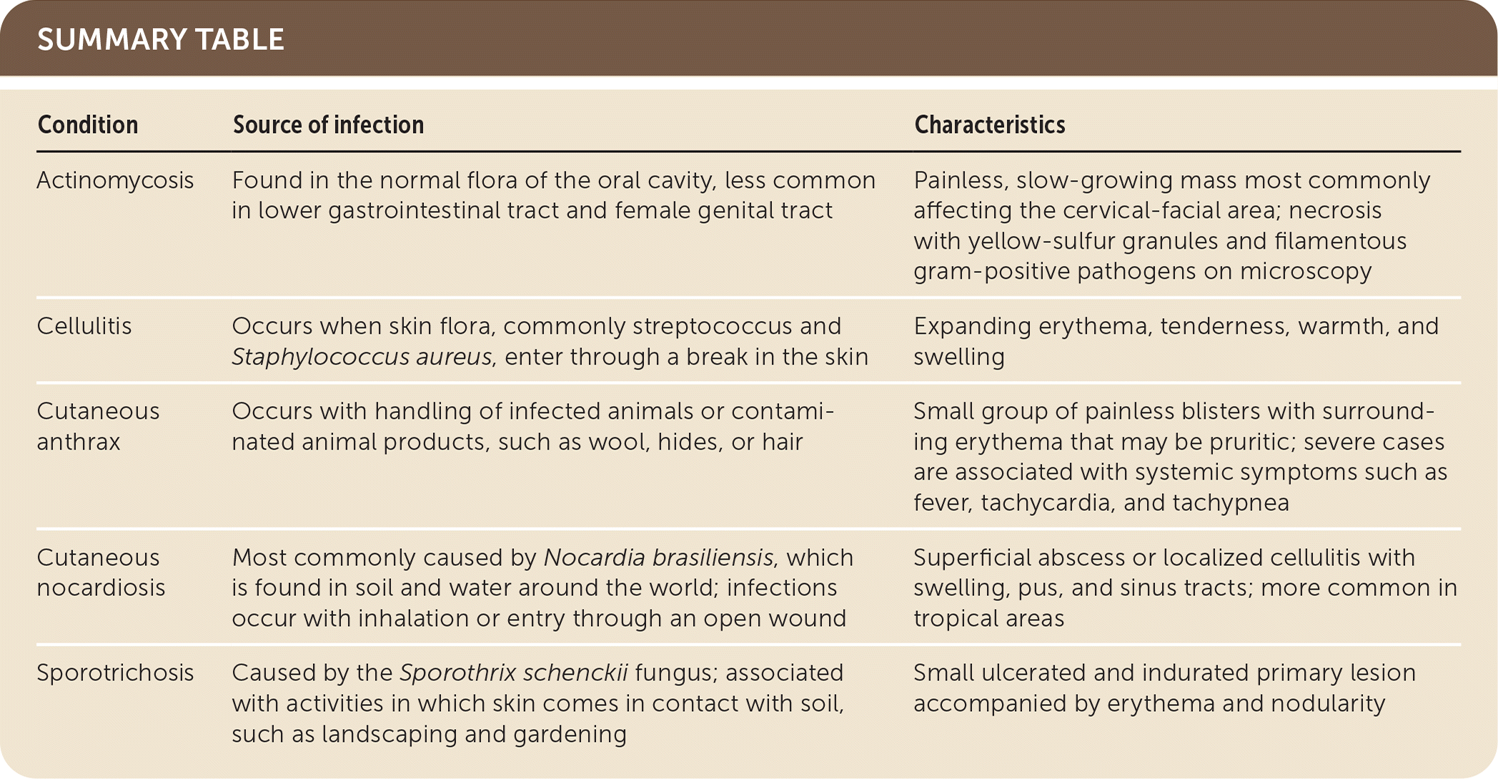
Answer is E: sporotrichosis, which is caused by the Sporothrix schenckii fungus. Sporotrichosis is a subacute to chronic infection associated with activities in which skin comes in contact with soil, such as landscaping and gardening.1 The most common clinical feature of lymphocutaneous sporotrichosis is a small ulcerated and indurated primary lesion accompanied by erythema and nodularity.2 Similar lesions appear proximal to the primary lesion, along the lymphatic channels. On initial presentation, pain is usually mild and systemic symptoms absent. Biopsy and fungal culture are the diagnostic standards for sporotrichosis.3
Actinomycosis is a rare disease caused by Actinomyces, an anaerobic gram-positive species found in the normal oral flora and less commonly in the lower gastrointestinal tract and female genital tract. It sometimes spreads to other parts of the body through breaks in the mucous membranes and devitalized tissue, causing illness. Actinomycosis usually presents as a painless, slow-growing mass most commonly affecting the cervical-facial area. Typical microscopic findings are necrosis with yellow-sulfur granules and filamentous gram-positive pathogens.4
Cellulitis is a soft tissue infection that occurs when skin flora enters through a break in the skin. It presents as expanding erythema, tenderness, warmth, and swelling. Because most cases are nonculturable, an organism is not usually identified. When an organism is isolated, beta-hemolytic streptococcus and Staphylococcus aureus are most common.5
Cutaneous anthrax is a zoonotic disease caused by Bacillus anthracis. It is the most common form of human anthrax. Mild cases are characterized by a small group of painless blisters with surrounding erythema that can be pruritic, with a lack of systemic symptoms. Severe cases are notable for a bullous eruption presenting as a large lesion associated with extensive edema and systemic symptoms such as fever, tachycardia, and tachypnea.6
Cutaneous nocardiosis is most commonly caused by Nocardia brasiliensis, which is found in soil and water around the world. Infection occurs with inhalation or entry through an open wound and is more common in tropical areas. Cutaneous nocardiosis manifests as a superficial abscess or localized cellulitis with swelling, pus, and sinus tracts. It can spread to the regional lymph nodes and produce a chain of nodular lesions. Mycetoma, a painless mass usually on the foot, can develop in advanced disease.7
| Condition | Source of infection | Characteristics |
|---|---|---|
| Actinomycosis | Found in the normal flora of the oral cavity, less common in lower gastrointestinal tract and female genital tract | Painless, slow-growing mass most commonly affecting the cervical-facial area; necrosis with yellow-sulfur granules and filamentous gram-positive pathogens on microscopy |
| Cellulitis | Occurs when skin flora, commonly streptococcus and Staphylococcus aureus, enter through a break in the skin | Expanding erythema, tenderness, warmth, and swelling |
| Cutaneous anthrax | Occurs with handling of infected animals or contaminated animal products, such as wool, hides, or hair | Small group of painless blisters with surrounding erythema that may be pruritic; severe cases are associated with systemic symptoms such as fever, tachycardia, and tachypnea |
| Cutaneous nocardiosis | Most commonly caused by Nocardia brasiliensis, which is found in soil and water around the world; infections occur with inhalation or entry through an open wound | Superficial abscess or localized cellulitis with swelling, pus, and sinus tracts; more common in tropical areas |
| Sporotrichosis | Caused by the Sporothrix schenckii fungus; associated with activities in which skin comes in contact with soil, such as landscaping and gardening | Small ulcerated and indurated primary lesion accompanied by erythema and nodularity |
