
A more recent article on acute headache in adults is available.
Am Fam Physician. 2013;87(10):682-687
Related editorial: Hard-to-Diagnose Headache: Practical Tips for Diagnosis and Treatment
Author disclosure: No relevant financial affiliations.
Approximately one-half of the adult population worldwide is affected by a headache disorder. The International Headache Society classification and diagnostic criteria can help physicians differentiate primary headaches (e.g., tension, migraine, cluster) from secondary headaches (e.g., those caused by infection or vascular disease). A thorough history and physical examination, and an understanding of the typical features of primary headaches, can reduce the need for neuroimaging, lumbar puncture, or other studies. Some red flag signs and symptoms identified in the history or during a physical examination can indicate serious underlying pathology and will require neuroimaging or other testing to evaluate the cause of headache. Red flag signs and symptoms include focal neurologic signs, papilledema, neck stiffness, an immunocompromised state, sudden onset of the worst headache in the patient's life, personality changes, headache after trauma, and headache that is worse with exercise. If an intracranial hemorrhage is suspected, head computed tomography without contrast media is recommended. For most other dangerous causes of headache, magnetic resonance imaging or computed tomography is acceptable.
Headache is a common pain condition worldwide. It is important for physicians evaluating adult patients with acute headache to determine whether the condition is benign or if it indicates dangerous neurologic or systemic pathology. The most common types of headaches are tension-type headaches, migraines, and cluster headaches, which affect approximately 40, 10, and 1 percent of the adult population, respectively.1,2
| Clinical recommendation | Evidence rating | References |
|---|---|---|
| A diagnosis of migraine is highly likely with presence of headache with nausea, or if the patient reports experiencing two of three features from either of these symptom triads: nausea, photophobia, or pulsating pain; or nausea, photophobia, or a headache that worsens with exertion. | C | 15 |
| Head computed tomography should be performed before lumbar puncture in all patients with suspected subarachnoid hemorrhage, regardless of findings on neurologic examination. | C | 23 |
| A patient with sudden onset of severe headache (e.g., patient reporting the worst headache of his or her life, or maximal from initiation, or thunderclap headache) should be evaluated with computed tomography of the head without contrast media. | C | 28 |
| Immunocompromised patients with severe headache should be evaluated with magnetic resonance imaging of the head with and without contrast media. | C | 28 |
Most headache diagnoses are based entirely on the patient history. Only rarely does physical examination provide clues to the diagnosis.3
The International Headache Society has published a system of classification and operational diagnostic criteria for headache based on clinical consensus.4 This system is most useful for classifying patients in epidemiologic studies and clinical trials. Classifying headaches into primary (tension, migraine, or cluster) and secondary types (e.g., those caused by infection or vascular disease) is also useful to differentiate headaches that, although perhaps recurrent and temporarily disabling, have no dangerous underlying cause from those that may be a sign of significant pathology, because they represent an underlying systemic or neurologic disorder (Table 1).5
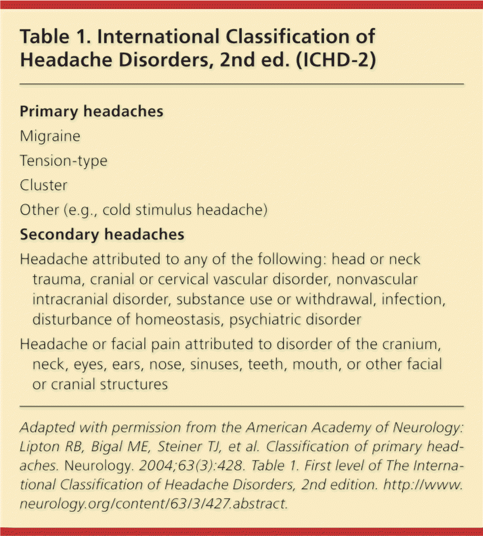
| Primary headaches |
| Migraine |
| Tension-type |
| Cluster |
| Other (e.g., cold stimulus headache) |
| Secondary headaches |
| Headache attributed to any of the following: head or neck trauma, cranial or cervical vascular disorder, nonvascular intracranial disorder, substance use or withdrawal, infection, disturbance of homeostasis, psychiatric disorder |
| Headache or facial pain attributed to disorder of the cranium, neck, eyes, ears, nose, sinuses, teeth, mouth, or other facial or cranial structures |
Primary Headaches
Patients with a history of headache who do not have red flag signs and symptoms are at low risk of serious headache. Additionally, they should have primary headache characteristics (Tables 1 through 5).4,5 Criteria for low-risk headaches are listed in Table 6.6 Patients at low risk of serious headache do not require neuroimaging.7
TENSION-TYPE HEADACHE
Tension-type headache is the most common form of headache, and affects more than 40 percent of the adult population worldwide.1 It is characterized by bilateral mild to moderate pressure without other associated symptoms.4 Women are affected slightly more often than men.8 Nociceptors in the pericranial myofascial tissues are a likely source of tension headaches.9,10 Several studies have found that individuals who experience chronic tension-type headaches have increased sensitivity to pressure, electrical stimuli, and thermal stimuli in the pericranial myofascial tissue, and can find even normally harmless stimuli painful.10–12 Individuals who meet the criteria for tension-type headache but who have normal neurologic examination results require no additional laboratory testing or neuroimaging.13 Classification criteria for tension-type headaches are listed in Table 2.5
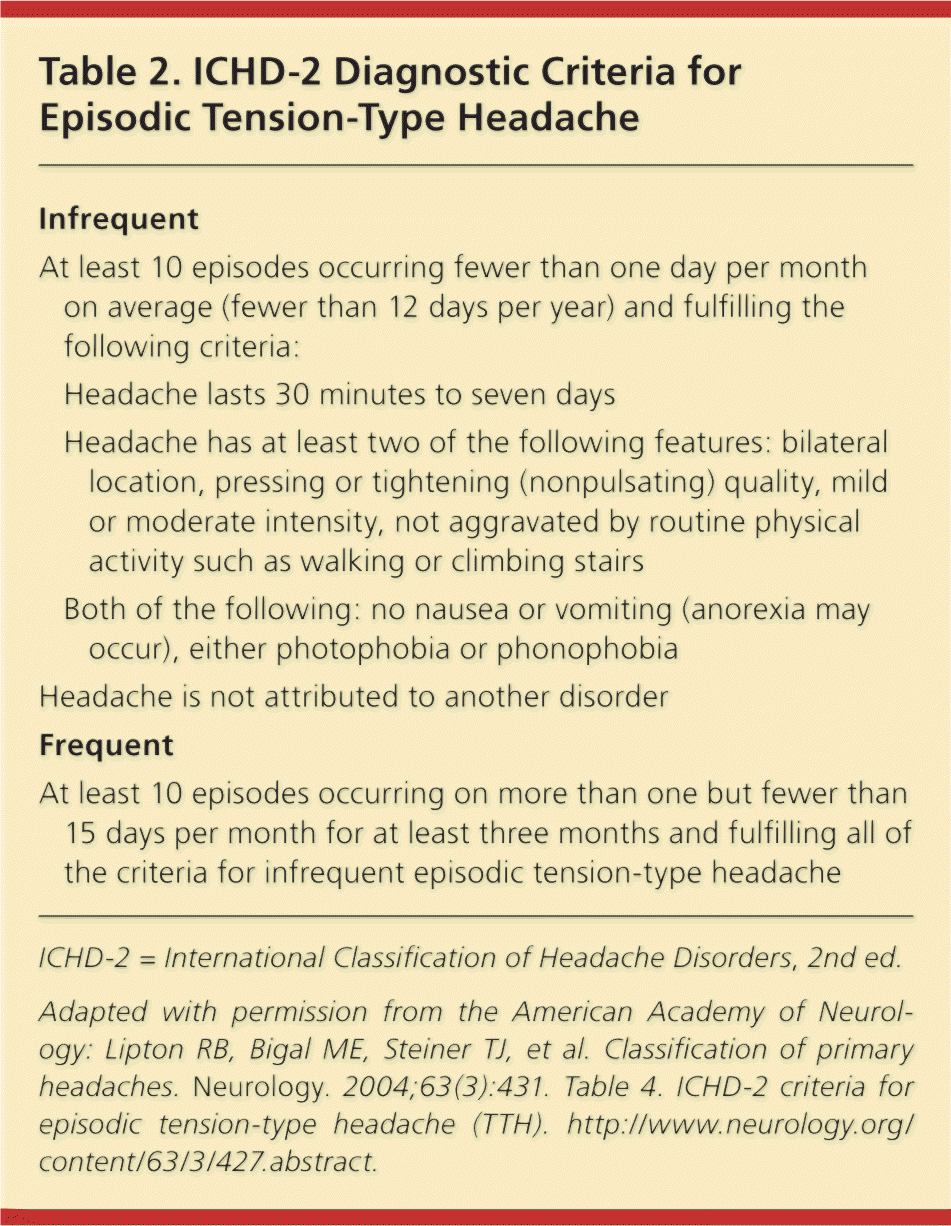
| Infrequent | |
| At least 10 episodes occurring fewer than one day per month on average (fewer than 12 days per year) and fulfilling the following criteria: | |
| Headache lasts 30 minutes to seven days | |
| Headache has at least two of the following features: bilateral location, pressing or tightening (nonpulsating) quality, mild or moderate intensity, not aggravated by routine physical activity such as walking or climbing stairs | |
| Both of the following: no nausea or vomiting (anorexia may occur), either photophobia or phonophobia | |
| Headache is not attributed to another disorder | |
| Frequent | |
| At least 10 episodes occurring on more than one but fewer than 15 days per month for at least three months and fulfilling all of the criteria for infrequent episodic tension-type headache | |
MIGRAINE HEADACHES
Useful clinical criteria from the history and physical examination for distinguishing migraine from tension-type headache include nausea, photophobia (sensitivity to light), and phonophobia (sensitivity to sound). Physical activity often exacerbates migraine headache. Combined findings useful for distinguishing migraine can be summarized by the POUND mnemonic (pulsatile quality, duration of four to 72 hours, unilateral location, nausea or vomiting, and disabling intensity). Patients who meet at least four of these criteria are most likely to have a migraine.14
One study of 1,500 adults with migraine headache found that the presence of nausea alone, or the presence of two of three features from either of these symptom triads (i.e., nausea, photophobia, and pulsating quality; or nausea, photophobia, and worsening of headache with physical activity) had positive likelihood ratios for migraine of 4.8 or greater and negative likelihood ratios of less than 0.23.15
| At least two episodes fulfilling the following criteria: | |
| Aura consisting of at least one of the following, but no motor weakness: fully reversible visual symptoms including positive features (e.g., flickering lights, spots or lines) and/or negative features (i.e., loss of vision); fully reversible sensory symptoms including positive features (i.e., pins and needles) and/or negative features (i.e., numbness); fully reversible dysphasic speech disturbance | |
| At least two of the following: homonymous visual symptoms and/or unilateral symptoms; at least one aura symptom develops gradually over five or more minutes and/or different aura symptoms occur in succession over five or more minutes; each symptom lasts at least five minutes, but no longer than 60 minutes | |
| A headache that fulfills the criteria for migraine without aura (Table 4), and begins during the aura or follows the aura within 60 minutes | |
| Headache not attributed to another disorder | |
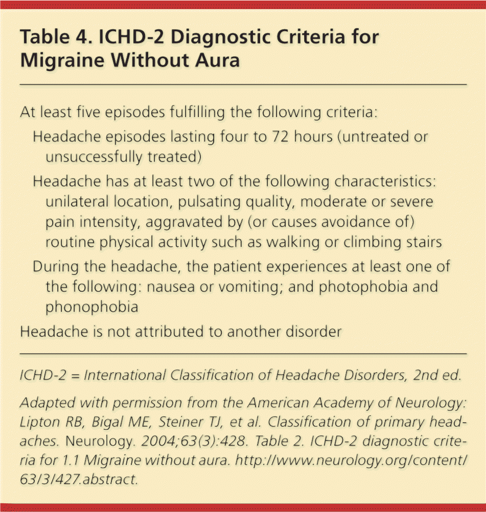
| At least five episodes fulfilling the following criteria: | |
| Headache episodes lasting four to 72 hours (untreated or unsuccessfully treated) | |
| Headache has at least two of the following characteristics: unilateral location, pulsating quality, moderate or severe pain intensity, aggravated by (or causes avoidance of) routine physical activity such as walking or climbing stairs | |
| During the headache, the patient experiences at least one of the following: nausea or vomiting; and photophobia and phonophobia | |
| Headache is not attributed to another disorder | |
CLUSTER HEADACHES
Cluster headaches are relatively rare, and are characterized by brief (15 to 180 minutes) episodes of severe head pain with associated autonomic symptoms1 (Table 54 ). Although cluster headaches are less common than migraines and tension-type headaches, an estimated 500,000 Americans experience them at least once in a lifetime.16 The age of onset of cluster headaches varies, with 70 percent of patients reporting onset before 30 years of age.17
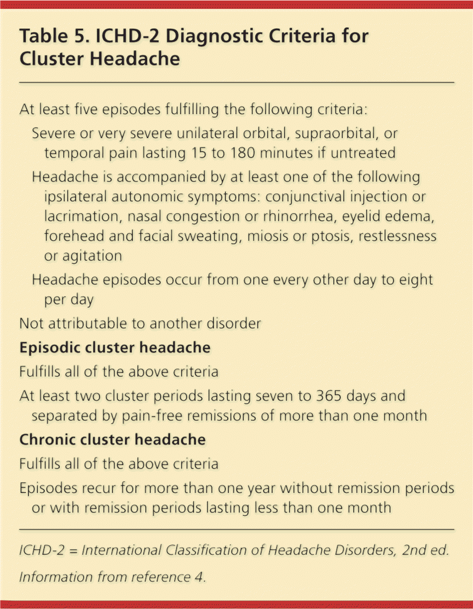
| At least five episodes fulfilling the following criteria: | |
| Severe or very severe unilateral orbital, supraorbital, or temporal pain lasting 15 to 180 minutes if untreated | |
| Headache is accompanied by at least one of the following ipsilateral autonomic symptoms: conjunctival injection or lacrimation, nasal congestion or rhinorrhea, eyelid edema, forehead and facial sweating, miosis or ptosis, restlessness or agitation | |
| Headache episodes occur from one every other day to eight per day | |
| Not attributable to another disorder | |
| Episodic cluster headache | |
| Fulfills all of the above criteria | |
| At least two cluster periods lasting seven to 365 days and separated by pain-free remissions of more than one month | |
| Chronic cluster headache | |
| Fulfills all of the above criteria | |
| Episodes recur for more than one year without remission periods or with remission periods lasting less than one month | |
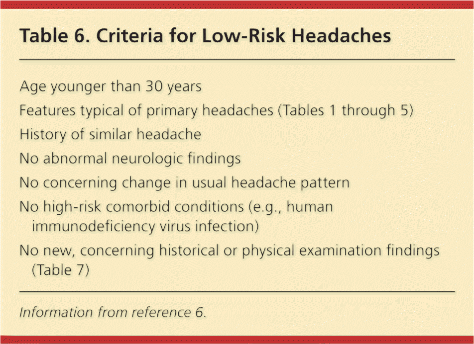
| Age younger than 30 years |
| Features typical of primary headaches (Tables 1 through 5) |
| History of similar headache |
| No abnormal neurologic findings |
| No concerning change in usual headache pattern |
| No high-risk comorbid conditions (e.g., human immunodeficiency virus infection) |
| No new, concerning historical or physical examination findings (Table 7) |
Patients with cluster headache most commonly describe the pain as sharp, but some report that it can also be pulsating and pressure-like. Although pain can occur on both sides of the head, most patients report unilateral pain. Pain most commonly occurs in the retro-orbital area, followed by the temporal region, upper teeth, jaw, cheek, lower teeth, and neck.17 Ipsilateral autonomic symptoms such as eyelid edema, nasal congestion, lacrimation, or forehead sweating usually accompany the pain. There tend to be several (up to eight) episodes in the same day, with each episode lasting between 15 and 180 minutes.4 In the episodic form (80 to 90 percent of cases), episodes occur daily for a number of weeks followed by a period of remission.4 On average, a period of cluster headaches lasts six to 12 weeks, with remission lasting up to 12 months.4 In the chronic form (10 to 20 percent of cases), episodes occur without significant periods of remission.4
The long delay in diagnosis reported by patients who have cluster headaches is important. Only 25 percent of patients with cluster headaches are diagnosed correctly within one year of symptom onset, and more than 40 percent report a delay in diagnosis of five years or longer.16 The most common incorrect diagnoses reported in one study were migraine (34 percent), sinusitis (21 percent), and allergies (6 percent).15 Family history appears to have a role in some cases. A number of comorbidities are associated with cluster headaches, including depression (24 percent), sleep apnea (14 percent), restless legs syndrome (11 percent), and asthma (9 percent).15 Depression is an important diagnosis, because many individuals who have cluster headaches report suicidal thoughts, and 2 percent of patients in one study had attempted suicide.16,18,19
Dangerous Headaches
Distinguishing dangerous headaches from benign or low-risk headaches is a significant challenge because the symptoms can overlap. Recommendations for differentiating dangerous from benign headaches are provided in Table 7.5,20–24 The characteristics of dangerous headaches and associated red flag symptoms are based on observational study and consensus reports. Therefore, they are not absolutely accurate in identifying serious underlying causes in patients who have headache.
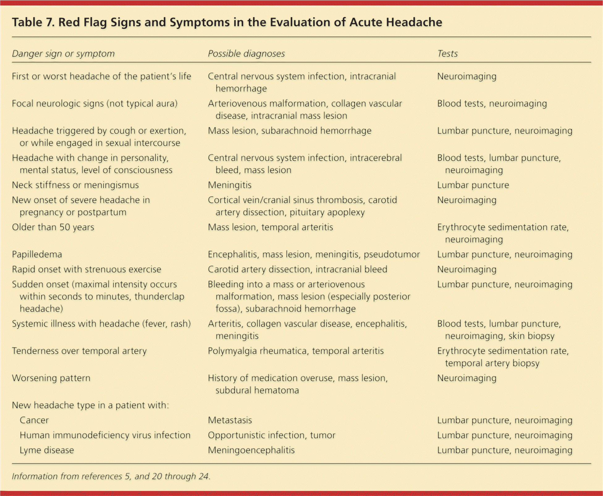
| Danger sign or symptom | Possible diagnoses | Tests | |
|---|---|---|---|
| First or worst headache of the patient's life | Central nervous system infection, intracranial hemorrhage | Neuroimaging | |
| Focal neurologic signs (not typical aura) | Arteriovenous malformation, collagen vascular disease, intracranial mass lesion | Blood tests, neuroimaging | |
| Headache triggered by cough or exertion, or while engaged in sexual intercourse | Mass lesion, subarachnoid hemorrhage | Lumbar puncture, neuroimaging | |
| Headache with change in personality, mental status, level of consciousness | Central nervous system infection, intracerebral bleed, mass lesion | Blood tests, lumbar puncture, neuroimaging | |
| Neck stiffness or meningismus | Meningitis | Lumbar puncture | |
| New onset of severe headache in pregnancy or postpartum | Cortical vein/cranial sinus thrombosis, carotid artery dissection, pituitary apoplexy | Neuroimaging | |
| Older than 50 years | Mass lesion, temporal arteritis | Erythrocyte sedimentation rate, neuroimaging | |
| Papilledema | Encephalitis, mass lesion, meningitis, pseudotumor | Lumbar puncture, neuroimaging | |
| Rapid onset with strenuous exercise | Carotid artery dissection, intracranial bleed | Neuroimaging | |
| Sudden onset (maximal intensity occurs within seconds to minutes, thunderclap headache) | Bleeding into a mass or arteriovenous malformation, mass lesion (especially posterior fossa), subarachnoid hemorrhage | Lumbar puncture, neuroimaging | |
| Systemic illness with headache (fever, rash) | Arteritis, collagen vascular disease, encephalitis, meningitis | Blood tests, lumbar puncture, neuroimaging, skin biopsy | |
| Tenderness over temporal artery | Polymyalgia rheumatica, temporal arteritis | Erythrocyte sedimentation rate, temporal artery biopsy | |
| Worsening pattern | History of medication overuse, mass lesion, subdural hematoma | Neuroimaging | |
| New headache type in a patient with: | |||
| Cancer | Metastasis | Lumbar puncture, neuroimaging | |
| Human immunodeficiency virus infection | Opportunistic infection, tumor | Lumbar puncture, neuroimaging | |
| Lyme disease | Meningoencephalitis | Lumbar puncture, neuroimaging | |
Patients with characteristics of secondary headache should be evaluated to determine whether the headache is dangerous. Computed tomography of the head is the most widely used imaging study for acute head trauma because of its availability, speed, and accuracy. However, magnetic resonance imaging of the brain is more sensitive for detecting subdural hematoma, and is therefore particularly important in identifying smaller lesions.20
An algorithm for diagnosing headaches is available from the Institute for Clinical Systems Improvement at https://www.icsi.org/_asset/qwrznq/Headache.pdf.3
HISTORY AND PHYSICAL EXAMINATION
History. Thunderclap headache, which is characterized by sudden-onset headache pain, with peak intensity occurring within several minutes, requires prompt evaluation. Subarachnoid hemorrhage, hypertensive emergencies, vertebral artery dissections, and acute angle–closure glaucoma can also present this way.25
Use of illicit drugs, including cocaine and methamphetamine, can increase the risk of intracranial bleeding or stroke. Prescription or over-the-counter medications such as aspirin, other nonsteroidal anti-inflammatory drugs, anticoagulants, and glucocorticoids increase the risk of intracranial bleeding.
A history of human immunodeficiency virus infection or other immunosuppressive conditions in patients with headache may suggest a brain abscess, meningitis, or malignancy of the central nervous system (CNS).21,26 The presence of a coexisting infection in the lungs, sinuses, or orbital areas may precede and cause a CNS infection.
A patient who reports the worst headache of his or her life, especially if the patient is older than 50 years, or who has a headache that occurs with exertion (including sexual intercourse) could be experiencing intracranial hemorrhage or carotid artery dissection.26 Prompt investigation is required for any headaches associated with neurologic findings, including changes in mental status, seizures, and visual disturbances. Additional red flag symptoms and signs are listed in Table 7.5,20–24
Physical Examination. Neurologic abnormalities require evaluation and are particularly concerning in association with acute headache. Abnormalities are one of the best predictors of CNS pathology.6,14,27 A focal neurologic deficit should not be attributed to migraine headache unless a similar pattern has occurred with a previous migraine. By definition, aura associated with migraine lasts 60 minutes or less. Therefore, headache with aura-like symptoms should not be assumed to be benign or a primary headache when aura-like symptoms are present for more than 60 minutes.
Abnormal findings on examination can be pronounced, such as meningismus or unilateral vision loss, or subtle, such as extensor plantar response or unilateral pronator drift. Obtundation or confusion suggests a dangerous headache because these signs do not occur with benign or primary headache.
Patients with headache and fever, papilledema, or severe hypertension (systolic pressure greater than 180 mm Hg or diastolic pressure greater than 120 mm Hg) require evaluation for CNS infection and increased intracranial pressure. Patients also should be evaluated to determine if their blood pressure should be lowered to safer levels to avoid intracranial hemorrhage from malignant hypertension. Contusions and facial or scalp lacerations increase the likelihood of associated intracranial hemorrhage (Table 75,20–24 ).
DIAGNOSTIC TESTING
Neuroimaging. Neuroimaging is indicated for all patients who present with signs or symptoms of dangerous headache, because they are at increased risk of intracranial pathology. Although considerable debate exists about the optimal way to perform neuroimaging for acute headaches, the American College of Radiology has made a few specific recommendations (Table 8).28
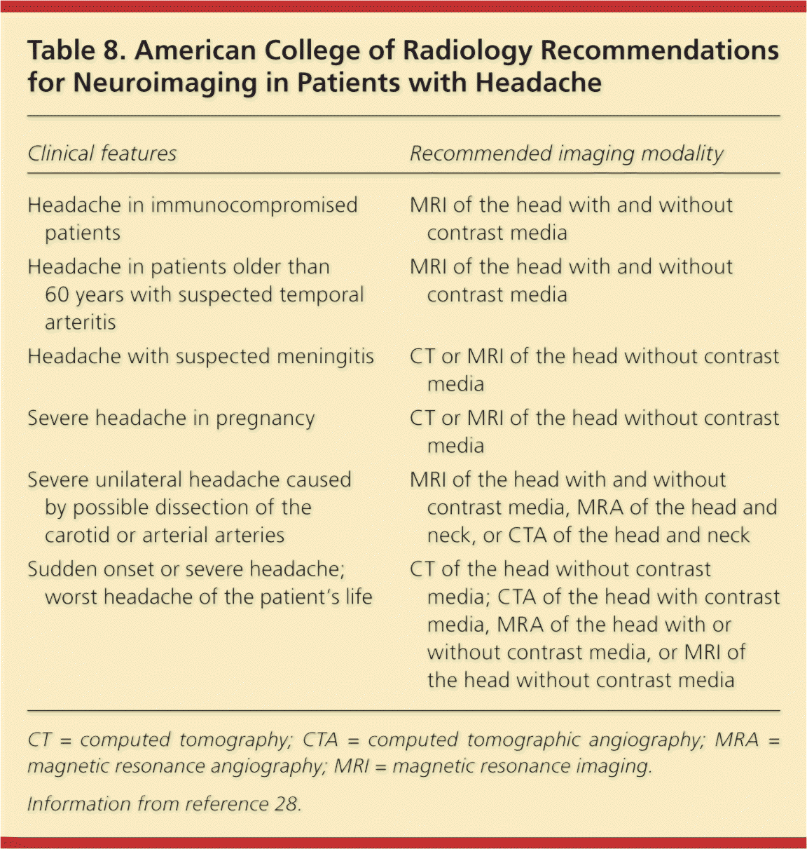
| Clinical features | Recommended imaging modality |
|---|---|
| Headache in immunocompromised patients | MRI of the head with and without contrast media |
| Headache in patients older than 60 years with suspected temporal arteritis | MRI of the head with and without contrast media |
| Headache with suspected meningitis | CT or MRI of the head without contrast media |
| Severe headache in pregnancy | CT or MRI of the head without contrast media |
| Severe unilateral headache caused by possible dissection of the carotid or arterial arteries | MRI of the head with and without contrast media, MRA of the head and neck, or CTA of the head and neck |
| Sudden onset or severe headache; worst headache of the patient's life | CT of the head without contrast media; CTA of the head with contrast media, MRA of the head with or without contrast media, or MRI of the head without contrast media |
Lumbar Puncture. Lumbar puncture is useful for identifying infection, the presence of red blood cells (which suggests bleeding), and abnormal cells associated with some CNS malignancies. In adults with suspected subarachnoid hemorrhage, it is important to perform lumbar puncture to check for blood or xanthochromia. Computed tomography of the head should be performed before lumbar puncture, even if the results of neurologic examination are normal, because there is a risk of central herniation of the brain even in the absence of physical examination findings of subarachnoid hemorrhage. In one supporting study, 5 percent of patients presenting to an emergency department with suspected subarachnoid hemorrhage and a normal neurologic examination had early intracranial herniation or midline shift.29
Response to Pain Relief. The American College of Emergency Physicians has determined that response to pain relief therapy should not be used as the sole diagnostic indicator of the underlying etiology of an acute headache.13 No prospective randomized controlled trials, evidence from meta-analyses, randomized controlled trials, or well-designed cohort studies support or refute the practice of using response to pain relief therapy in nontraumatic headaches as an indicator of potential underlying pathology.
Data Sources: We performed a PubMed search for headache topics, and reviewed recent relevant publications in the Cochrane database, Essential Evidence Plus, and the National Guideline Clearinghouse. The search included expert consensus statements, clinical reviews, and clinical trials. Search terms included headache, acute headache, and classification of headache. Search date: December 2011.
