Corneal abrasions result from cutting, scratching, or abrading the thin, protective, clear coat of the exposed anterior portion of the ocular epithelium. These injuries cause pain, tearing, photophobia, foreign body sensation, and a gritty feeling. Symptoms can be worsened by exposure to light, blinking, and rubbing the injured surface against the inside of the eyelid. Visualizing the cornea under cobalt-blue filtered light after the application of fluorescein can confirm the diagnosis. Most corneal abrasions heal in 24 to 72 hours and rarely progress to corneal erosion or infection. Although eye patching traditionally has been recommended in the treatment of corneal abrasions, multiple well-designed studies show that patching does not help and may hinder healing. Topical mydriatics also are not beneficial. Initial treatment should be symptomatic, consisting of foreign body removal and analgesia with topical non-steroidal anti-inflammatory drugs or oral analgesics; topical antibiotics also may be used. Corneal abrasions can be avoided through the use of protective eyewear.
Most of the human eye lies within a protective bony orbit. The exposed anterior portion has other anatomic and functional protections. The eyebrow and eyelashes partially shield the eye from small particles. Eyelids close rapidly and reflexively when ocular danger is sensed. A tear response attempts to wash away anything that reaches the ocular surface. Tears also lubricate the eye and prevent tissue dehydration.
Strength of Recommendation (SOR) Labels
| Key clinical recommendations | SOR labels | References |
|---|---|---|
| Patching is not effective for treatment of corneal abrasions and is not recommended. | A | 2, 3, 6 |
| Consider topical nonsteroidal anti-inflammatory drugs in patients with corneal abrasions. | A | 9 |
| Topical mydriatics are not effective for treatment of corneal abrasions and are not recommended. | B | 10 |
| Consider use of topical antibiotics in patients with corneal abrasions. | C | 11, 12 |
| Discontinue contact lens use in patients with corneal abrasions. | C | 13 |
Despite built-in protections, eye injuries still occur. One such injury is abrasion of the outermost layer of the eye. Although damage to the white part of the eye usually is of little significance, corneal abrasion can be serious. When minor abrasions occur, healthy cells quickly fill the defect to prevent vision-diminishing infection or irregularity in refraction. If the abrasion penetrates the cornea more deeply, the healing process takes longer—24 to 72 hours.1,2 Deeper scratches can cause corneal scarring that can impair vision to the point where corneal transplant is needed. Specific incidence and prevalence data are not available, but corneal abrasion is the most common eye injury in children presenting to emergency departments.3
Function and Structure of the Cornea
The cornea (Figure 1) is a highly organized group of cells and proteins with three functions: barrier protection, filtration of some of the ultraviolet wavelengths in sunlight, and refraction (the cornea is responsible for 65 to 75 percent of the eye’s capacity to focus light on the retina). The cornea must be totally transparent to refract light properly. Therefore, it has no blood vessels and instead is nourished by tears, environmental oxygen, and the aqueous humor of the anterior chamber.
Figure 1
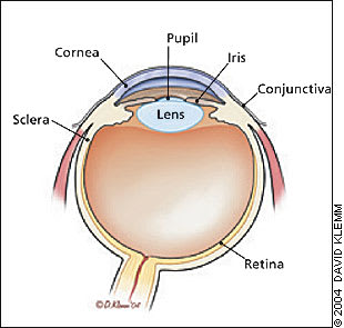
Cornea in relationship to the rest of the eye.
Figure 2
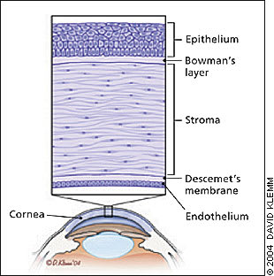
Anatomy of the cornea.
Within its thin dimensions—about 11.6 mm vertically, 10.5 mm horizontally, 1 mm thick peripherally, and 0.55 mm thick centrally—the cornea has five distinct, transparent layers; from anterior to posterior they are epithelium, Bowman’s layer, stroma, Descemet’s membrane, and endothelium (Figure 2).
Diagnosing Corneal Abrasion
A history of recent ocular trauma and subsequent acute pain suggests corneal abrasion. Other symptoms include photophobia, pain with extraocular muscle movement, excessive tearing, blepharospasm, foreign body sensation, gritty feeling, blurred vision, and headache. Symptoms can be present without the patient’s recollection of trauma and with as little trauma as aggressive eye rubbing.
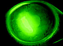
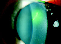
The diagnosis of corneal abrasion can be confirmed by visualizing the cornea under cobalt-blue filtered light after the application of fluorescein, which will cause the abrasion to appear green (Figures 3 and 4) . If examination is limited by pain, instillation of a topical anesthetic (e.g., proparacaine [Ophthetic], tetracaine [Pontocaine]) may be needed. During the examination it is important to assess for and remove any foreign bodies, some of which may leave a rust residue (Figure 5) .
Figure 3
Corneal abrasion stained with fluorescein.
Figure 4
Corneal abrasion stained with fluorescein and highlighted by cobalt blue light.
Figure 5
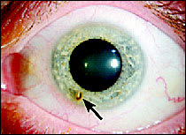
rust ring remaining after removal of a small, metallic foreign body.
Rarely, simple corneal abrasions become complicated. Recurrent corneal erosion (RCE)—repeated, spontaneous disruption of corneal epithelium—can occur in corneal tissue weakened by abrasion months or years earlier. Symptoms of RCE include ocular pain, foreign body sensation, photophobia, blepharospasm, decreased vision, and lacrimation on awakening or after rubbing or opening the eyes. These symptoms are annoying to the patient but typically are not severe enough to interfere with activities.4 Lesions usually are found near the original abrasion; they may recur only rarely or as often as daily. True idiopathic or bilateral lesions suggest a basement membrane dystrophy, characterized by poor adhesion between the epithelial basement membrane and Bowman’s layer.
Treatment Options
Although eye patches, topical antibiotics, and mydriatic agents traditionally have been used in patients with corneal abrasions, treatment recommendations recently have evolved. Current recommendations stress the use of topical or oral analgesics and topical antibiotics (Table 1) . Most corneal abrasions heal with this approach.
TABLE 1 Topical NSAIDs and Antibiotics
| Drug | Dosage | Price (generic)* | Comments | |
|---|---|---|---|---|
| Topical NSAIDs | ||||
| Diclofenac (Voltaren), 0.1% solution | One drop four times daily | $52 for 5 mL | May delay wound healing. Use caution in patients withbleeding tendencies. | |
| Ketorolac (Acular), 0.5% solution | One drop four times daily | $56 for 5 mL | Avoid use in patients whowear contact lenses. Discontinue use if epithelium breakdown occurs. | |
| Topical antibiotics | ||||
| Bacitracin (AK-Tracin), 500 units per g ointment | 1/2-inch ribbon two to four times daily | $5 for 3.5 g | ||
| Chloramphenicol (Chloroptic), 1% ointment | Two drops every three hours | $22 for 3.5 g | Discontinue use if no improvement after one week. | |
| Ciprofloxacin (Ciloxan), 0.3% solution | Day 1: two drops every 15 minutes for six hours, then two drops every 30 minutes for rest of day | |||
| Day 2: Two drops per hour | ||||
| Days 3 to 14: Two drops every four hours | $45 for 5 mL | Anti-pseudomonal activity | ||
| Erythromycin 0.5% ointment | 1/2-inch ribbon two to four times daily | $3 to $6 for 3.5 g | ||
| Gentamycin (Garamycin), 0.3% ointment or solution | One to two drops every four hours or 1/2-inch ribbon two to three times daily | $10 ($5 to $10) for 5 mL | Anti-pseudomonal activity | |
| Ofloxacin (Ocuflox), 0.3% solution | Days 1 and 2: One to two drops every 30 minutes | |||
| Days 3 to 7: One to two drops per hour | ||||
| Day 8 to treatment completion: One to two drops four times daily. | $40 for 5 mL | Anti-pseudomonal activity | ||
NSAID = nonsteroidal anti-inflammatory drug
*— Estimated cost to the pharmacist based on average wholesale prices in Red book. Montvale, N.J.: Medical Economics Data, 2004. Cost to the patient will be higher, depending on prescription filling fee.
EYE PATCHING
Eye patching is no longer recommended for corneal abrasions.2,3,5 A meta-analysis of five randomized controlled trials (RCTs) failed to reveal an increase in healing rate or improvement on a pain scale.5 Two subsequent RCTs (one in children, one in adults) reported similar results.2,3 In the past, patching was thought to reduce pain by reducing blinking and decreasing eyelid-induced trauma to the damaged cornea. However, the patch itself was the main cause of pain in 48 percent of patients.6 Children with patches had greater difficulty walking than those without patches.3 Furthermore, patching can result in decreased oxygen delivery, increased moisture, and a higher chance of infection. Thus, patching may actually retard the healing process.7,8
TOPICAL ANALGESICS
Topical nonsteroidal anti-inflammatory drugs (NSAIDs) such as diclofenac (Voltaren) and ketorolac (Acular) are modestly useful in reducing pain from corneal abrasions.9 In a systematic review of five RCTs, topical NSAID use decreased pain by an average of 1.3 cm on a standard 10-cm pain scale.9 Qualitatively, patients using topical NSAIDs indicated greater relief from pain and other symptoms.9 Patients using topical NSAIDs may take fewer oral analgesics (two of three studies), return to work earlier (one study), and require fewer narcotics.9
Topical anesthetics should be avoided after the initial examination. They can retard healing and cause corneal damage.
MYDRIATICS
Mydriatics are no longer recommended for the treatment of pain in patients with corneal abrasions.10 Mydriatics formerly were prescribed to relieve ciliary muscle spasm that was thought to occur in patients with corneal abrasions. However, in one RCT with limited follow-up, pain was similar in patients using an eye lubricant or mydriatic (2 percent homatropine [Homapin]), alone or combined with a topical NSAID.10
TOPICAL ANTIBIOTICS
Because a concomitant infection can cause slower healing of corneal abrasions, some clinicians use prophylactic antibiotic treatment, although there is no strong evidence for this use. A two-year, non–placebo-controlled, prospective cohort study11 of topical antibiotic prophylaxis for corneal abrasion showed that the use of 1 percent chloramphenicol ointment was associated with lower risk of subsequent ulcer, especially if prophylaxis began within 18 hours after the injury. A single-blind, non–placebo-controlled randomized trial12 showed that corneal abrasions in patients treated with fusidic acid eye drops did not heal significantly faster than patients treated with chloramphenicol ointment.
If antibiotics are used, ointment (e.g., baci-tracin [AK-Tracin], erythromycin, gentamycin [Garamycin]) is more lubricating than drops and is considered first-line treatment. In patients who wear contact lenses, an anti-pseudomonal antibiotic (e.g., ciprofloxacin [Ciloxan], gentamycin, ofloxacin [Ocuflox]) should be used, and contact lens use should be discontinued. Clinical trial data are lacking, but it is recommended that contact lenses be avoided until the abrasion is healed and the antibiotic course completed.13
ORAL ANALGESICS
No direct evidence is available from clinical trials for the efficacy of oral analgesics in the treatment of corneal abrasions. However, because most abrasions heal without significant long-term complications, pain relief is the primary concern and the basis for routine use of oral analgesics. Oral analgesics are less expensive than topical preparations. No studies directly address the role, if any, of opioid analgesia. Individual patient characteristics (e.g., age, concomitant illness, drug allergy, ability to tolerate NSAIDs, potential for opioid abuse, employment conditions such as driving and machine operation) should guide therapy.
Follow-up and Referral Guidelines
Most patients should be re-evaluated in 24 hours; if the abrasion has not fully healed, they should be evaluated again three to four days later. Patients who wear contact lenses should be re-evaluated in 24 hours and again three to four days later even if they feel well. Any worsening of symptoms should prompt a thorough re-evaluation for foreign bodies or full-thickness injuries. Immunocompromised or monocular patients also warrant closer attention and may require earlier ophthalmologic referral.
Referral to an ophthalmologist is indicated for patients with deep eye injuries, foreign bodies that cannot be removed, and suspected RCE. Patients with persistent symptoms after three days, worsening symptoms, and symptoms that do not improve daily also should be referred. Patients who wear contact lenses should be referred if there is no improvement in symptoms within a few hours of lens removal.
Primary Prevention and Screening
Most corneal abrasions are preventable. Persons in high-risk occupations (e.g., miners, woodworkers, metal workers, landscapers) and those who participate in certain sports (e.g., hockey, lacrosse, racquetball) should wear eye protection. Levels of protection include plastic safety glasses, polycarbonate lenses of varying thickness, industrial safety goggles with polycarbonate, and helmets with facemasks. All provide barrier protection from airborne debris (e.g., sand, sawdust, metal) and other objects that could cause ocular trauma (e.g., fingernails, tree branches, sports balls). Eye guards without lenses are not sufficient. Other preventive measures include careful fitting and placement of contact lenses, keeping the fingernails of infants and young children clipped short, and removing low-hanging tree branches or objects from the home environment.
Corneal abrasion, the most common peri-operative ocular injury, results from lagophthalmos during general anesthesia. It can be prevented by taping the patient’s eyelids closed or instilling soft contact lenses or aqueous gels; paraffin-based ointments (e.g., Lacrilube, Duratears) appear to be less effective.14
Screening is important in three populations: neonates on mask ventilation, sedated or paralyzed patients on a ventilator, and persons who wear contact lenses. Corneal abrasion, with subsequent Pseudomonas panophthalmitis, can occur in patients in neonatal intensive care units who are receiving continuous positive airway pressure ventilation. It is attributed to the pressure of the masks on the orbit.15 Eye discharge in mask-ventilated neonates should prompt evaluation for corneal abrasion and infection.
A similar problem can occur in adults who are deeply sedated or receiving neuromuscular blocking agents while on a ventilator, because their protective corneal reflex is suppressed. The incidence of corneal abrasion in this population decreased from 18 to 4 percent when prophylactic lubricating ointment was administered every four hours.16 Persons who wear contact lenses are at higher risk of developing abrasions that become infected and ulcerate (Figure 6). Soft, extended-wear lenses have been associated with a 10-fold to 15-fold increase in ulcerative keratitis.17 Case reports and a nonsystematic review suggest that screening for corneal abrasions also may be needed after airbag deployment in automobile crashes.18,19
Figure 6
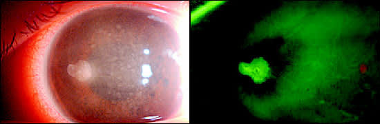
Corneal ulcer in a patient who wears contact lenses. (Left) View without fluorescein stain. (Right) View with fluorescein stain.
Prognosis
Healing time depends on the size of the corneal abrasion. Most abrasions heal in two to three days, while larger abrasions that involve more than one half of the surface area of the cornea may take four to five days.1 In patients with traumatic corneal abrasions who are treated in ophthalmology offices, 28 percent had recurrent symptoms up to three months after the injury.4
