Ischemic stroke is the third leading cause of death in the United States and a common reason for hospitalization. The subacute period after a stroke refers to the time when the decision to not employ thrombolytics is made up until two weeks after the stroke occurred. Family physicians are often involved in the subacute management of ischemic stroke. All patients with an ischemic stroke should be admitted to the hospital in the subacute period for cardiac and neurologic monitoring. Imaging studies, including magnetic resonance angiography, carotid artery ultrasonography, and/or echocardiography, may be indicated to determine the cause of the stroke. Evaluation for aspiration risk, including a swallowing assessment, should be performed, and nutritional, physical, occupational, and speech therapy should be initiated. Significant causes of morbidity and mortality following ischemic stroke include venous thromboembolism, pressure sores, infection, and delirium, and measures should be taken to prevent these complications. For secondary prevention of future strokes, antiplatelet therapy with aspirin should be initiated within 24 hours of ischemic stroke in all patients without contraindications, and one of several antiplatelet regimens should be continued long-term. Statin therapy should also be given in most situations. Although permissive hypertension is initially warranted, antihypertensive therapy should begin within 24 hours. Diabetes mellitus should be controlled and patients counseled about lifestyle modifications to reduce stroke risk. Rehabilitative therapy following hospitalization improves outcomes and should be considered.
Ischemic stroke, which includes thrombotic and embolic subtypes, is a major cause of morbidity and mortality worldwide. In the United States, it is the third leading cause of death and is a common reason for hospitalization.1 Despite significant advances in the acute therapies for ischemic stroke (i.e., thrombolytics and mechanical clot disruption or retrieval), less than 5 percent of patients qualify for these interventions mainly because they do not present early enough.2,3 Risk factors for ischemic stroke are listed in Table 1.
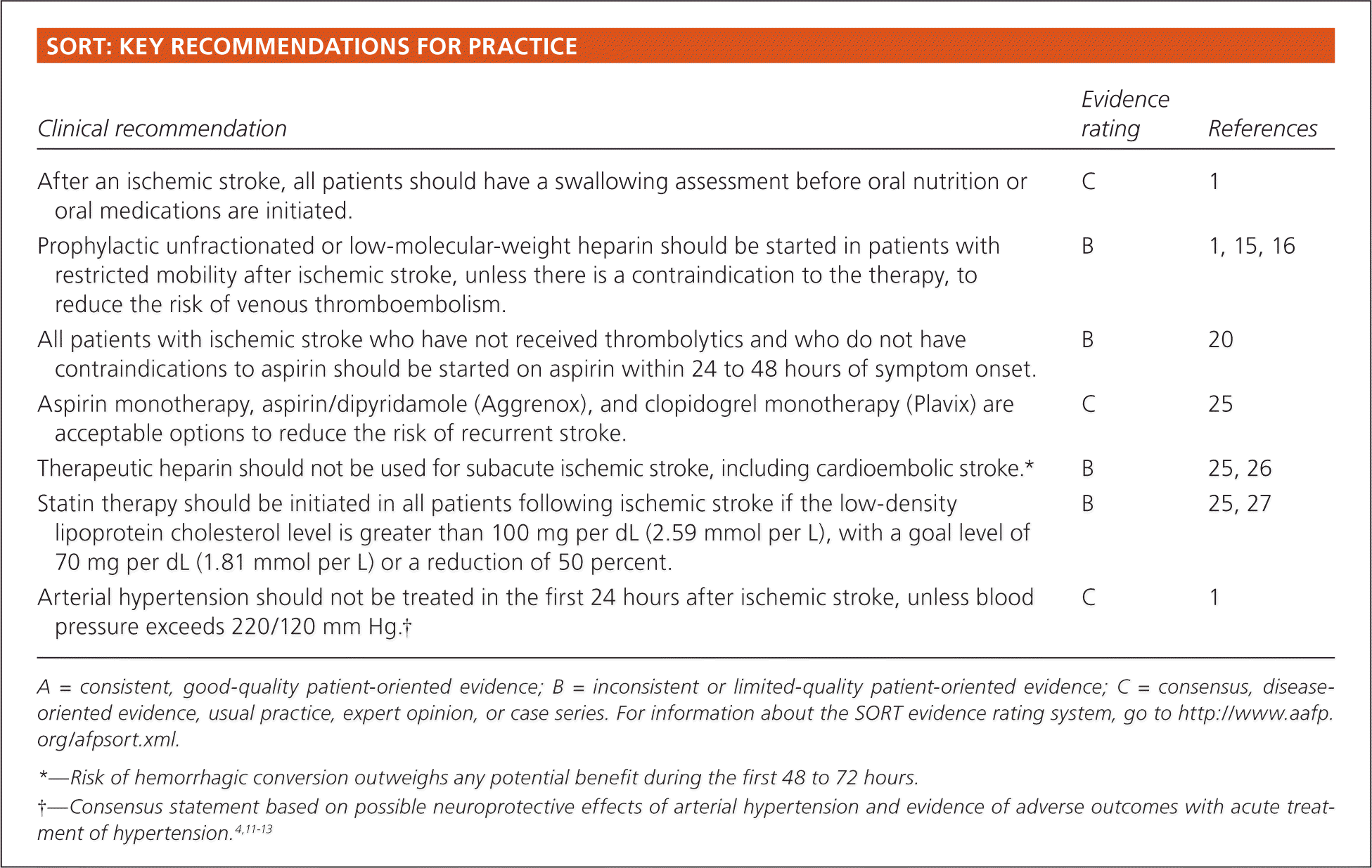
SORT: KEY RECOMMENDATIONS FOR PRACTICE
| Clinical recommendation | Evidence rating | References |
|---|---|---|
| After an ischemic stroke, all patients should have a swallowing assessment before oral nutrition or oral medications are initiated. | C | 1 |
| Prophylactic unfractionated or low-molecular-weight heparin should be started in patients with restricted mobility after ischemic stroke, unless there is a contraindication to the therapy, to reduce the risk of venous thromboembolism. | B | 1, 15, 16 |
| All patients with ischemic stroke who have not received thrombolytics and who do not have contraindications to aspirin should be started on aspirin within 24 to 48 hours of symptom onset. | B | 20 |
| Aspirin monotherapy, aspirin/dipyridamole (Aggrenox), and clopidogrel monotherapy (Plavix) are acceptable options to reduce the risk of recurrent stroke. | C | 25 |
| Therapeutic heparin should not be used for subacute ischemic stroke, including cardioembolic stroke.* | B | 25, 26 |
| Statin therapy should be initiated in all patients following ischemic stroke if the low-density lipoprotein cholesterol level is greater than 100 mg per dL (2.59 mmol per L), with a goal level of 70 mg per dL (1.81 mmol per L) or a reduction of 50 percent. | B | 25, 27 |
| Arterial hypertension should not be treated in the first 24 hours after ischemic stroke, unless blood pressure exceeds 220/120 mm Hg† | C | 1 |
A = consistent, good-quality patient-oriented evidence; B = inconsistent or limited-quality patient-oriented evidence; C = consensus, disease-oriented evidence, usual practice, expert opinion, or case series. For information about the SORT evidence rating system, go to https://www.aafp.org/afpsort.xml.
*—Risk of hemorrhagic conversion outweighs any potential benefit during the first 48 to 72 hours.
†—Consensus statement based on possible neuroprotective effects of arterial hypertension and evidence of adverse outcomes with acute treatment of hypertension.4,11–13
Table 1. Risk Factors for Ischemic Stroke (Embolic or Thrombotic)
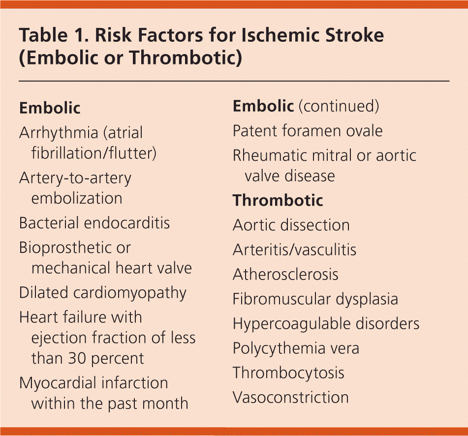
| Embolic |
| Arrhythmia (atrial fibrillation/flutter) |
| Artery-to-artery embolization |
| Bacterial endocarditis |
| Bioprosthetic or mechanical heart valve |
| Dilated cardiomyopathy |
| Heart failure with ejection fraction of less than 30 percent |
| Myocardial infarction within the past month |
| Embolic |
| Patent foramen ovale |
| Rheumatic mitral or aortic valve disease |
| Thrombotic |
| Aortic dissection |
| Arteritis/vasculitis |
| Atherosclerosis |
| Fibromuscular dysplasia |
| Hypercoagulable disorders |
| Polycythemia vera |
| Thrombocytosis |
| Vasoconstriction |
Subacute management of ischemic stroke refers to the period from when the decision to not employ thrombolytics is made up until two weeks after the stroke occurred. Family physicians are often involved in the care of patients during the subacute period. Careful subacute treatment of patients in the hospital and later in outpatient settings is critical to preventing unnecessary complications, recurrent stroke, and death.
All patients presenting with an ischemic stroke should be admitted to the hospital.1 The goals of hospital care are listed in Table 2. Thrombolytic therapy can be considered for up to six hours after symptom onset, and physicians with expertise in these therapies should be consulted for management assistance.2,3 Patients who have been treated with thrombolytics should be admitted to an intensive care unit skilled in the management of postthrombolytic care. This article focuses on subacute treatment of patients who do not receive thrombolytics.
Table 2. Goals of Hospital Care After Ischemic Stroke
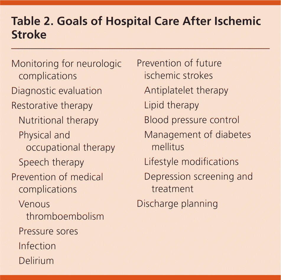
| Monitoring for neurologic complications | |
| Diagnostic evaluation | |
| Restorative therapy | |
| Nutritional therapy | |
| Physical and occupational therapy | |
| Speech therapy | |
| Prevention of medical complications | |
| Venous thromboembolism | |
| Pressure sores | |
| Infection | |
| Delirium | |
| Prevention of future ischemic strokes | |
| Antiplatelet therapy | |
| Lipid therapy | |
| Blood pressure control | |
| Management of diabetes mellitus | |
| Lifestyle modifications | |
| Depression screening and treatment | |
| Discharge planning | |
Monitoring for Neurologic Complications
Following an ischemic stroke, patients are at risk of multiple neurologic complications, including cerebral edema, seizures, intracerebral hemorrhage (i.e., hemorrhagic conversion of an ischemic stroke), and evolution of neurologic deficits. Of these complications, cerebral edema is the leading cause of death in these patients.4
All patients initially should be assigned to cardiac monitoring units, with neurologic assessment throughout the first 24 to 48 hours. Although the optimal frequency of neurologic assessment has not been established, speech, extremity strength, and facial symmetry should be tested at least every two to three hours. Any worsening of neurologic status should prompt rapid evaluation with computed tomography (CT) to identify cerebral edema or hemorrhage, as well as strong consideration for consultation with a neurologist. Patients who have had a large cerebral or cerebellar ischemic stroke are at particularly high risk of serious morbidity and may require neurosurgical intervention; therefore, these patients should be transferred to a facility with 24-hour neurosurgical coverage.1
Diagnostic Evaluation
Patients admitted to the hospital following a stroke will have undergone diagnostic imaging with non–contrast-enhanced CT in the emergency department. After admission, the need for further imaging is based on whether the results would be beneficial in the treatment of the patient.
Diffusion-weighted magnetic resonance imaging of the brain can be useful for determining infarction size and confirming the presence of ischemic stroke if the diagnosis is in question (cerebral infarction often is not seen initially on CT imaging). Diffusion-weighted magnetic resonance imaging is the most sensitive and specific modality for early detection of acute cerebral infarction.5
Magnetic resonance angiography is useful for characterization of vascular disease. It has higher sensitivity and specificity than ultrasonography for detecting carotid artery disease. In addition to measuring atherosclerotic carotid stenosis (a common cause of ischemic stroke), it may also help identify less common etiologies, such as arterial dissection, venous thrombosis, fibromuscular dysplasia, and vasculitis. However, magnetic resonance angiography is significantly more expensive than ultrasonography, and its use is limited in patients who have metallic implants, pacemakers, or severe claustrophobia, or who cannot tolerate contrast media.6
Carotid artery ultrasonography is an inexpensive and useful modality for identifying atherosclerotic carotid stenosis.6 Patients with greater than 70 percent carotid stenosis on the side of the neurologic deficit may have a reduced risk of future stroke if carotid endarterectomy is performed, preferably within two weeks of the ischemic event.7,8 Patients with moderate ipsilateral stenosis (50 to 69 percent) may benefit from early endarterectomy, depending on patient-specific factors (e.g., age, sex, comorbidity, severity of initial symptoms), but the evidence is less clear. Surgical intervention has not been shown to benefit patients with ipsilateral carotid stenosis of less than 50 percent.9
There are no clear recommendations regarding the routine use of echocardiography in the evaluation of ischemic stroke. Although it is commonly performed in the initial diagnostic workup, its value may be limited when there is low suspicion for an embolic source. In patients with risk factors for embolic stroke (Table 1), some studies have shown that transthoracic echocardiography alone can miss up to 32 percent of pertinent abnormalities, such as intracardiac thrombi that would require anticoagulation or structural lesions (e.g., patent foramen ovale) that may benefit from surgical repair.10 If a patient has any risk factor for embolic stroke and no pertinent abnormality is detected on transthoracic echocardiography, transesophageal echocardiography is recommended.
Restorative Therapies
NUTRITIONAL THERAPY
Aspiration is often a preventable complication of stroke. Evaluation for risk factors leading to aspiration, including a swallowing assessment, should be performed during the initial examination of a patient with subacute stroke, before initiating oral nutrition or oral medications.1 Oral intake should be restricted in patients at risk of aspiration because of obtundation or an obvious swallowing dysfunction until a swallowing assessment is performed by a properly trained health care professional. A modified barium swallow test may be necessary in some patients.11
Measures should be taken to avoid dehydration and malnutrition because both lead to poor outcomes after a stroke.12 All patients who are unable to take nutrition or fluids orally should receive adequate intravenous maintenance fluids and enteral feedings via nasogastric or nasoduodenal tubes. A percutaneous endoscopic gastrostomy can be considered if prolonged enteral nutrition is necessary and the patient (or the health care proxy if the patient is unable to communicate) desires it.
PHYSICAL AND OCCUPATIONAL THERAPY
Physical and occupational therapy are important components of recovery for patients with stroke and should not be delayed.13 Early initiation can improve functional outcomes and reduce the incidence of deep vein thrombosis, contracture, pressure sores, and pneumonia.1
SPEECH THERAPY
Not all patients with subacute stroke will have a speech deficit. However, for those who do, there is some evidence that early intensive speech therapy improves outcomes.14
Prevention of Medical Complications
VENOUS THROMBOEMBOLISM
Unfractionated heparin and low-molecular-weight heparin have been shown to reduce the incidence of venous thromboembolism after ischemic stroke.15 Evidence on sequential leg compression devices is lacking, but they are probably less effective than heparin-based products and should be used only when heparin is not an option.
Current guidelines recommend a prophylactic dose of unfractionated or low-molecular-weight heparin for patients with restricted mobility after ischemic stroke, unless there is a specific contraindication to the therapy.1,15,16 Fondaparinux (Arixtra) is also an option for the prevention of venous thromboembolism after stroke.17
PRESSURE SORES
Early mobilization, frequent turning and skin examinations, and use of alternating pressure mattresses should be considered to prevent skin damage after ischemic stroke.13 Assessing nutritional status with measurement of albumin, prealbumin, and cholesterol levels can aid in identifying patients at higher risk of wound formation (low levels indicate higher risk).
INFECTION
Patients with neurologic deficits leading to immobility are at higher risk of pneumonia from atelectasis and aspiration. These patients should be mobilized early in the hospital stay and encouraged to use bedside incentive spirometry hourly to aid deep breathing and ease cough. Medications that reduce the level of consciousness or increase risk of aspiration pneumonia (e.g., proton pump inhibitors) should be discontinued unless specifically indicated.18 Urinary tract infections are common after stroke, and indwelling catheters should be avoided. Fever is independently linked to poor outcomes in patients with stroke. Although antipyretics have not been shown to improve neurologic outcomes, a workup for the source of any fever should be performed.1
DELIRIUM
Delirium, defined as an acute change in cognition and attention that fluctuates throughout the day, can develop after stroke, especially in older adults.19 Delirium can lead to increased length of hospitalization, high death rates, and limited functional capacity. Measures to reduce the risk of delirium after stroke include limiting anticholinergic and sedative medications, avoiding disturbances of the sleep-wake cycle, and preserving available sensory input.19
Prevention of Future Ischemic Strokes
ANTIPLATELET THERAPY
Antiplatelet therapy is a cornerstone of the secondary prevention of ischemic stroke. There is consensus that all patients who have not received thrombolytics and who do not have contraindications to aspirin should be given aspirin within 24 to 48 hours of symptom onset.20 The optimal regimen for longer-term antiplatelet therapy is unclear despite multiple studies comparing antiplatelet medications20; key studies are summarized in Table 3.21–24
Table 3. Key Studies Comparing Antiplatelet Medications for Secondary Prevention of Ischemic Stroke
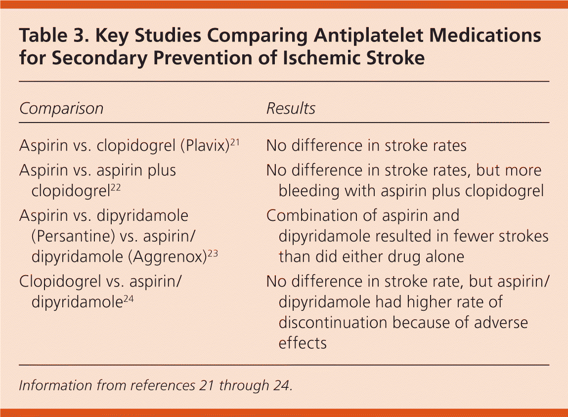
| Comparison | Results |
|---|---|
| Aspirin vs. clopidogrel (Plavix)21 | No difference in stroke rates |
| Aspirin vs. aspirin plus clopidogrel22 | No difference in stroke rates, but more bleeding with aspirin plus clopidogrel |
| Aspirin vs. dipyridamole (Persantine) vs. aspirin/ dipyridamole (Aggrenox)23 | Combination of aspirin and dipyridamole resulted in fewer strokes than did either drug alone |
| Clopidogrel vs. aspirin/dipyridamole24 | No difference in stroke rate, but aspirin/dipyridamole had higher rate of discontinuation because of adverse effects |
In 2011, the American Heart Association (AHA) and the American Stroke Association (ASA) issued new guidelines on secondary prevention of ischemic stroke.25 According to the guidelines, aspirin monotherapy (50 to 325 mg per day), aspirin combined with extended-release dipyridamole (Aggrenox), and clopidogrel monotherapy (Plavix) are all acceptable regimens for secondary prevention of ischemic stroke. The guidelines do not provide specific recommendations about second-line agents for patients already on antiplatelet therapy.
In the past, patients received anticoagulation with heparin during the subacute period, but more recent studies have not shown benefit from this approach.26 The AHA/ASA guidelines recommend against administering heparin (other than at low doses for prevention of venous thromboembolism) in the subacute management of ischemic stroke, including cardioembolic stroke, because the risk of hemorrhagic conversion outweighs any potential benefit during the first 48 to 72 hours.25
LIPID THERAPY
The lipid-lowering agent atorvastatin (Lipitor) has been evaluated for secondary prevention of ischemic stroke.27 The study, which compared 80 mg of atorvastatin (initiated 30 days after the stroke) with placebo, showed a statistically significant decrease in stroke recurrence in patients taking atorvastatin compared with those taking placebo. Although an optimal dosage or drug choice is not specified, the AHA/ASA guidelines recommend that a statin be prescribed for patients following an atherosclerotic ischemic stroke if the low-density lipoprotein level is greater than 100 mg per dL (2.59 mmol per L).25 For these patients and those already on a statin, the guidelines recommend a goal of 70 mg per dL (1.81 mmol per L) or a reduction of 50 percent.
BLOOD PRESSURE CONTROL
Elevated blood pressure may have a protective effect in the initial period after an ischemic stroke, and studies have shown adverse outcomes when blood pressure is lowered in the acute period.28 Most patients who present with hypertension during a stroke will have a spontaneous decrease in blood pressure, without any treatment, in the first several hours after presentation.28 Therefore, it is prudent not to treat elevated blood pressure for 24 hours after an acute stroke, unless the blood pressure exceeds 220/120 mm Hg or treatment is warranted by another medical condition, such as during an acute myocardial infarction.1
After 24 hours, tighter blood pressure control becomes more important for secondary prevention. In a post hoc analysis of the Perindopril Protection Against Recurrent Stroke Study (PROGRESS), patients with a blood pressure of 112/72 mm Hg or less had the lowest incidence of recurrent stroke.29 Another study demonstrated a 29 percent reduction in stroke recurrence with thiazide diuretics, even though the therapy reduced blood pressure by an average of only 5 mm Hg.30 The benefit of blood pressure reduction following an ischemic stroke for secondary prevention appears to extend to patients without hypertension.31
The AHA/ASA guidelines recommend that all patients receive antihypertensive therapy beginning 24 hours after symptom onset. There is insufficient evidence to make recommendations about the ideal blood pressure goal.25
DIABETES MANAGEMENT
Hyperglycemia is common in patients with or without diabetes mellitus who present with ischemic stroke,32 and studies have shown worse outcomes when the blood glucose level is elevated in the first 24 hours following the stroke.33 In most patients, therapy should be initiated when the blood glucose level is greater than 145 to 185 mg per dL (8.05 to 10.27 mmol per L).1 The optimal blood glucose goal for secondary prevention of ischemic stroke is unknown, and the AHA/ASA guidelines recommend a glycated hemoglobin goal that is consistent with current national diabetes guidelines.25
LIFESTYLE MODIFICATIONS
Smoking tobacco has been shown to double the risk of stroke, and even secondhand smoke raises the risk.34 Other important risk factors include excessive alcohol consumption, lack of physical activity, and obesity. Counseling regarding lifestyle modifications is critical following an ischemic stroke, and patients should receive direct counseling appropriate to their individual risk factors.25
DEPRESSION
In both the hospital and community settings, major depression ranges from 14.1 to 19.3 percent in patients who have had a stroke.35 Because depression has a negative influence on recovery, screening and appropriate treatment are warranted.35
Discharge Planning
There is strong evidence that use of rehabilitative services immediately following hospitalization benefits patients who have had a stroke, leading to better functional outcomes and increasing the odds of subsequently living at home.36 If available, transfer from the hospital to an inpatient rehabilitation unit should be strongly considered. If the patient's degree of disability does not warrant inpatient rehabilitation, outpatient therapy should be considered because it also reduces the odds of functional decline compared with no therapy.36
Data Sources: A PubMed search was performed using the search limits of meta-analysis, practice guideline, randomized controlled trial, review, English language, and published in the previous five years. Search terms were secondary prevention and ischemic stroke, acute management ischemic stroke, statin therapy and ischemic stroke, and blood pressure acute ischemic stroke. A Google-Scholar search was performed with the limit of articles published since 2001 and included citations. Search terms were secondary prevention ischemic stroke, ischemic stroke guidelines, and lipid control ischemic stroke. Search dates: July 2010 and May 2011.
