Am Fam Physician. 2023;108(1):70-77
Patient information: See related handout on stroke prevention.
Author disclosure: No relevant financial relationships.
Ischemic stroke is a major cause of morbidity and mortality worldwide. Ischemic stroke and transient ischemic attack exist on a continuum of the same disease process. Ischemic stroke is common, and more than 85% of stroke risk is attributed to modifiable risk factors. The initial management of acute stroke is usually performed in the emergency department and hospital settings. Family physicians have a key role in follow-up, ensuring that a complete diagnostic evaluation has been performed, addressing modifiable risk factors, facilitating rehabilitation, and managing chronic sequelae. Secondary prevention of ischemic stroke includes optimization of chronic disease management (e.g., hypertension, type 2 diabetes mellitus, dyslipidemia), nonpharmacologic lifestyle interventions (e.g., diet changes, exercise, substance use counseling), and pharmacologic interventions. Dual antiplatelet therapy with aspirin and clopidogrel is generally indicated for minor noncardioembolic ischemic strokes and high-risk transient ischemic attacks and should be converted to single antiplatelet therapy after 21 to 90 days. Secondary prevention of cardioembolic stroke requires long-term anticoagulation. Direct oral anticoagulants are preferred over warfarin for patients with nonvalvular atrial fibrillation. Poststroke problems with mobility, balance, cognition, dysphagia, and depression are common. Rehabilitation involves a multidisciplinary, multimodal approach that includes physical therapy, speech therapy, and treatment of chronic pain and poststroke depression.
Ischemic stroke is the result of a sudden blockage of blood flow to the central nervous system due to thrombotic or embolic phenomena, and it is a major cause of morbidity and mortality worldwide. In 2019, stroke was the fifth leading cause of death in the United States.1 Although stroke can be hemorrhagic or ischemic, approximately 87% of strokes in the United States are ischemic.2 Almost 800,000 strokes occur annually in the United States, often leading to long-term disability.2
Transient ischemic attack (TIA) is defined as a transient neurologic dysfunction caused by ischemia without infarction on imaging. Because the subsequent risk of stroke after TIA is similar to that of an initial stroke, TIA is treated similarly to a minor, nondisabling stroke. In this article, TIA is included with ischemic stroke, except where noted.

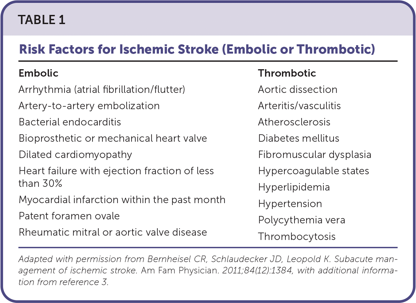
| Embolic Arrhythmia (atrial fibrillation/flutter) Artery-to-artery embolization Bacterial endocarditis Bioprosthetic or mechanical heart valve Dilated cardiomyopathy Heart failure with ejection fraction of less than 30% Myocardial infarction within the past month Patent foramen ovale Rheumatic mitral or aortic valve disease | Thrombotic Aortic dissection Arteritis/vasculitis Atherosclerosis Diabetes mellitus Fibromuscular dysplasia Hypercoagulable states Hyperlipidemia Hypertension Polycythemia vera Thrombocytosis |
Similar to many conditions, stroke is associated with health disparities. Compared with White people, Black people in the United States have strokes at younger ages and have more poststroke disability, much of which is related to modifiable risk factors.5 Increasing age and female sex are associated with greater poststroke disability.5 Stroke risk also varies with education level, income, zip code, health insurance, and social isolation, and assessing these factors is important for the primary and secondary prevention of stroke.6
Diagnostic Evaluation
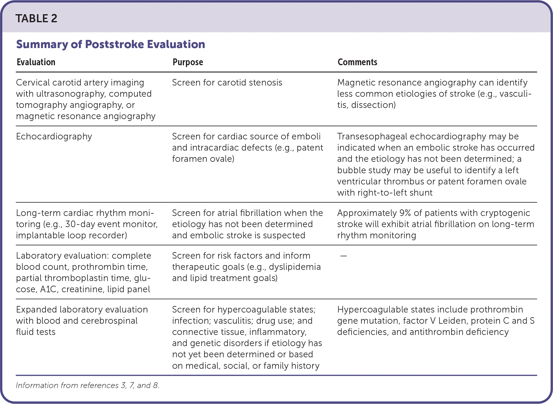
Although the initial management of acute stroke usually occurs in the emergency and hospital settings, the outpatient follow-up evaluation is critical to ensure that the appropriate testing has been completed, reviewed, and discussed with the patient.7 Certain parts of the evaluation to determine the cause of the stroke may be deferred until outpatient follow-up. Table 2 summarizes the evaluation of stroke on the basis of pathogenesis.3,7,8

| Evaluation | Purpose | Comments |
|---|---|---|
| Cervical carotid artery imaging with ultrasonography, computed tomography angiography, or magnetic resonance angiography | Screen for carotid stenosis | Magnetic resonance angiography can identify less common etiologies of stroke (e.g., vasculitis, dissection) |
| Echocardiography | Screen for cardiac source of emboli and intracardiac defects (e.g., patent foramen ovale) | Transesophageal echocardiography may be indicated when an embolic stroke has occurred and the etiology has not been determined; a bubble study may be useful to identify a left ventricular thrombus or patent foramen ovale with right-to-left shunt |
| Long-term cardiac rhythm monitoring (e.g., 30-day event monitor, implantable loop recorder) | Screen for atrial fibrillation when the etiology has not been determined and embolic stroke is suspected | Approximately 9% of patients with cryptogenic stroke will exhibit atrial fibrillation on long-term rhythm monitoring |
| Laboratory evaluation: complete blood count, prothrombin time, partial thromboplastin time, glucose, A1C, creatinine, lipid panel | Screen for risk factors and inform therapeutic goals (e.g., dyslipidemia and lipid treatment goals) | — |
| Expanded laboratory evaluation with blood and cerebrospinal fluid tests | Screen for hypercoagulable states; infection; vasculitis; drug use; and connective tissue, inflammatory, and genetic disorders if etiology has not yet been determined or based on medical, social, or family history | Hypercoagulable states include prothrombin gene mutation, factor V Leiden, protein C and S deficiencies, and antithrombin deficiency |
At the first primary care visit after stroke, diagnostic imaging should be reviewed to confirm the diagnosis.3 Although computed tomography (CT) and magnetic resonance imaging (MRI) are both used to diagnose stroke, MRI has 99% sensitivity for stroke, compared with 39% with CT, and can diagnose acute ischemic strokes missed by CT.8 If initial imaging was negative for stroke despite characteristic symptoms, follow-up CT or MRI may be indicated to confirm the diagnosis.3,7
Atrial fibrillation is estimated to cause approximately 1 in 7 strokes.9 In addition to electrocardiography performed during the initial evaluation of stroke, continuous cardiac rhythm monitoring is often performed during hospitalization.3 Long-term cardiac rhythm monitoring (e.g., 30-day event monitor, implantable loop recorder) increases the detection of atrial fibrillation and is useful in the evaluation of cryptogenic stroke.3,10
The initial evaluation of stroke includes laboratory testing to identify underlying risk factors and determine therapeutic targets.3 Complete blood count, prothrombin time, partial thromboplastin time, glucose, A1C, creatinine, and lipid profile are recommended to identify risk factors such as diabetes, dyslipidemia, chronic kidney disease, and blood cell dyscrasia. If stroke is determined to be cryptogenic based on the initial evaluation, expanded laboratory assessment, including testing for hypercoagulable states (e.g., prothrombin gene mutation, factor V Leiden, protein C and S deficiencies, antithrombin deficiency), infection, drug use, and connective tissue, inflammatory, and genetic disorders, may be indicated.
Polysomnography can also be considered to evaluate for obstructive sleep apnea. This condition is both a risk factor for stroke and a common comorbidity in patients with previous stroke (approximately 40% of stroke cases); however, it is unclear whether treatment with continuous positive airway pressure prevents recurrent vascular events.3
SECONDARY STROKE PREVENTION
Recurrent stroke risk is high, accounting for approximately 25% of all annual strokes in the United States, with the greatest risk of recurrence within the first week after a stroke.11 Numerous nonpharmacologic and pharmacologic strategies can help mitigate this risk.
LIFESTYLE MODIFICATIONS
Diets high in fat, processed meat, fried food, and sugar-sweetened beverages are associated with a significantly increased risk of stroke in adults.12 The American Heart Association/American Stroke Association (AHA/ASA) guidelines recommend a Mediterranean-style diet or a low-sodium Dietary Approaches to Stop Hypertension (DASH) diet for patients who have had a stroke.3,13 Increased physical activity is also recommended to prevent stroke recurrence.3
Although the recommendation for 150 minutes per week of moderate-intensity exercise applies to stroke survivors over the long-term, engaging in at least 10 minutes of moderate-intensity exercise four times per week, or 20 minutes of vigorous-intensity exercise two times per week, has been shown to reduce the risk of recurrent stroke. Breaking up sedentary time with three minutes of light activity every 30 minutes improves blood pressure.3,14
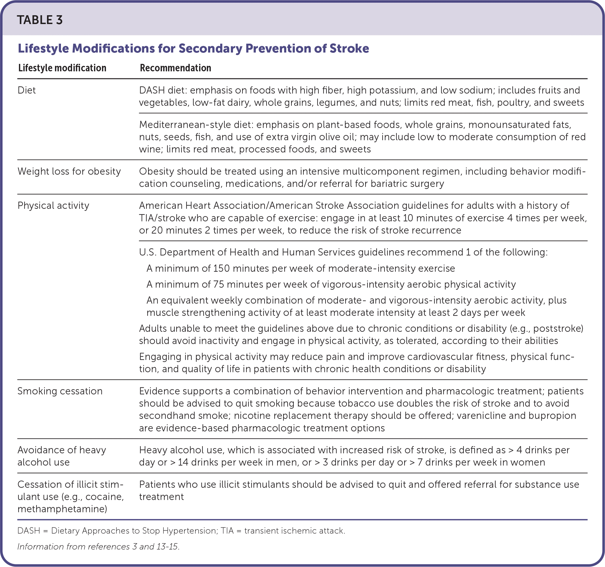
Weight loss is encouraged to improve comorbid hypertension, dyslipidemia, and type 2 diabetes. Substance use, including smoking, heavy alcohol use (more than four drinks per day for men or more than three drinks per day for women), and illicit stimulant use (e.g., cocaine, methamphetamine), should be addressed and treated. Table 3 details lifestyle recommendations after stroke.3,13–15

| Lifestyle modification | Recommendation |
|---|---|
| Diet | DASH diet: emphasis on foods with high fiber, high potassium, and low sodium; includes fruits and vegetables, low-fat dairy, whole grains, legumes, and nuts; limits red meat, fish, poultry, and sweets |
| Mediterranean-style diet: emphasis on plant-based foods, whole grains, monounsaturated fats, nuts, seeds, fish, and use of extra virgin olive oil; may include low to moderate consumption of red wine; limits red meat, processed foods, and sweets | |
| Weight loss for obesity | Obesity should be treated using an intensive multicomponent regimen, including behavior modification counseling, medications, and/or referral for bariatric surgery |
| Physical activity | American Heart Association/American Stroke Association guidelines for adults with a history of TIA/stroke who are capable of exercise: engage in at least 10 minutes of exercise 4 times per week, or 20 minutes 2 times per week, to reduce the risk of stroke recurrence |
| U.S. Department of Health and Human Services guidelines recommend 1 of the following: A minimum of 150 minutes per week of moderate-intensity exercise A minimum of 75 minutes per week of vigorous-intensity aerobic physical activity An equivalent weekly combination of moderate- and vigorous-intensity aerobic activity, plus muscle strengthening activity of at least moderate intensity at least 2 days per week Adults unable to meet the guidelines above due to chronic conditions or disability (e.g., poststroke) should avoid inactivity and engage in physical activity, as tolerated, according to their abilities Engaging in physical activity may reduce pain and improve cardiovascular fitness, physical function, and quality of life in patients with chronic health conditions or disability | |
| Smoking cessation | Evidence supports a combination of behavior intervention and pharmacologic treatment; patients should be advised to quit smoking because tobacco use doubles the risk of stroke and to avoid secondhand smoke; nicotine replacement therapy should be offered; varenicline and bupropion are evidence-based pharmacologic treatment options |
| Avoidance of heavy alcohol use | Heavy alcohol use, which is associated with increased risk of stroke, is defined as > 4 drinks per day or > 14 drinks per week in men, or > 3 drinks per day or > 7 drinks per week in women |
| Cessation of illicit stimulant use (e.g., cocaine, methamphetamine) | Patients who use illicit stimulants should be advised to quit and offered referral for substance use treatment |
TREATMENT OF COMORBID CEREBROVASCULAR RISK FACTORS
Hypertension is a major modifiable risk factor for stroke. The AHA/ASA guidelines recommend an ambulatory blood pressure goal of less than 130/80 mm Hg, if tolerated, for adults with a history of stroke.3 When choosing an anti-hypertensive medication, angiotensin-converting enzyme inhibitors, angiotensin receptor blockers, and thiazide diuretics have been shown to reduce the risk of recurrent stroke.3 Although calcium channel blockers are first-line treatment options for hypertension, their effectiveness for the secondary prevention of stroke is unproven.3
The AHA/ASA guidelines recommend an A1C goal of less than 7%.3 Intensive glycemic control has not been shown to improve the risk of recurrent stroke.3 Patients who are 65 years and older or have limited life expectancy may require less stringent A1C goals, consistent with American Diabetes Association recommendations.15 When possible, glucagon-like peptide-1 receptor agonists and sodium-glucose cotransporter-2 inhibitors should be considered to decrease the risk of cardiovascular events.3,15
The AHA/ASA guidelines recommend atorvastatin, 80 mg per day, or rosuvastatin (Crestor), 20 mg per day, to decrease the risk of recurrent stroke for patients with an atherosclerotic ischemic stroke and a low-density lipoprotein (LDL) cholesterol level greater than 100 mg per dL (2.59 mmol per L).3 For patients taking a statin, the LDL cholesterol goal is less than 70 mg per dL (1.81 mmol per L), regardless of history of coronary artery disease. If the LDL cholesterol goal cannot be achieved with statin monotherapy, the addition of ezetimibe or a proprotein convertase subtilisin/kexin type 9 inhibitor may be considered.3
ANTIPLATELET THERAPY
Indefinite antiplatelet therapy is indicated for secondary prevention of noncardioembolic ischemic stroke and cryptogenic stroke that do not appear to be embolic. Choice of antiplatelet drug depends on patient-specific factors such as tolerance, contraindications, presence of other vascular disease, availability, and cost.3 Aspirin, clopidogrel, and the combination of aspirin, 25 mg, and extended-release dipyridamole, 200 mg, are indicated for secondary prevention of ischemic stroke.
Short-term dual antiplatelet therapy can reduce the recurrence of some mild strokes.16,17 After minor noncardioembolic ischemic stroke with a National Institutes of Health Stroke Scale score of 3 or less (https://www.stroke.nih.gov/documents/NIH_Stroke_Scale_508C.pdf) or high-risk TIA with an ABCD2 score of 4 or greater (https://www.mdcalc.com/calc/357/abcd2-score-tia), the AHA/ASA guidelines recommend short-term treatment with aspirin, 81 mg per day, and clopidogrel, 75 mg per day, following initial loading doses of aspirin, 325 mg, and clopidogrel, 300 mg. Anti-platelet monotherapy or combination aspirin/dipyridamole therapy should be resumed indefinitely after 21 to 90 days.3 A shorter 10- to 21-day course of dual antiplatelet therapy before monotherapy may provide a similar benefit while minimizing bleeding risk.18 Use of dual antiplatelet therapy beyond 90 days confers no further risk reduction for prevention of recurrent stroke compared with single antiplatelet therapy but significantly increases bleeding risk.18
Long-term antiplatelet therapy regimens can include aspirin, 50 to 325 mg once per day; clopidogrel, 75 mg once per day; or combination aspirin, 25 mg, and extended-release dipyridamole, 200 mg, twice per day.19–21 Triple antiplatelet therapy should be avoided.3 It is unclear whether patients already taking aspirin at the time of noncardioembolic stroke or TIA benefit from increasing the dosage or changing to a different antiplatelet therapy.3
ANTICOAGULATION
After a stroke associated with atrial fibrillation, anticoagulation reduces future stroke risk by two-thirds and should be initiated unless contraindicated.9 For atrial fibrillation not associated with moderate mitral stenosis or a mechanical heart valve (e.g., nonvalvular atrial fibrillation), direct oral anticoagulants, such as apixaban (Eliquis), dabigatran (Pradaxa), edoxaban (Savaysa), or rivaroxaban (Xarelto), are preferred to vitamin K antagonists, such as warfarin, because of similar effectiveness and reduced bleeding risk.3,22
Patients with atrial fibrillation associated with moderate mitral stenosis or a mechanical heart valve have historically been excluded from trials involving direct oral anticoagulants; therefore, warfarin is recommended.3,22,23 Warfarin is recommended for patients with cardioembolic stroke secondary to thrombus of the left ventricle, left atrium, or left atrial appendage.3
TREATING CAROTID STENOSIS
In patients with single-sided extracranial internal carotid stenosis of at least 70%, carotid endarterectomy is recommended if perioperative risk is less than 6%. The short-term preoperative risk is later balanced by a reduction in recurrence, with a number needed to treat of 7 to prevent a recurrent stroke over five years.3 Surgery within two weeks of stroke is recommended over delayed surgery for optimal stroke prevention. Endarterectomy may be beneficial with lower levels of stenosis. Carotid artery stenting may be a reasonable alternative to endarterectomy for some patients.
Stroke Complications
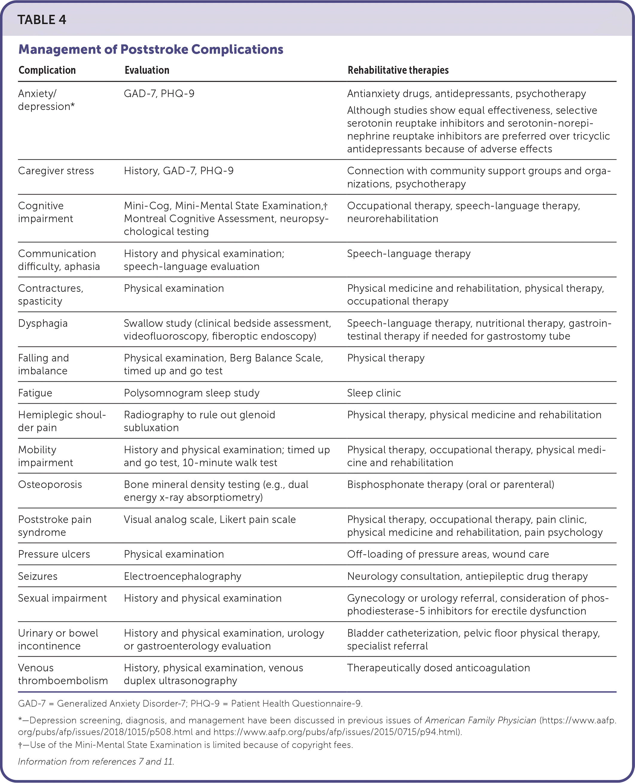
Stroke complications can include contractures, aphasia, hemiparesis, cognitive impairment, depression, anxiety, poststroke pain syndrome, headaches, and fatigue.7 Posthospitalization follow-up visits should focus on eliciting patient and caregiver experiences regarding the stroke, subsequent limitations, and unmet needs.7 Table 4 summarizes management of common stroke complications.7,11

| Complication | Evaluation | Rehabilitative therapies |
|---|---|---|
| Anxiety/depression* | GAD-7, PHQ-9 | Antianxiety drugs, antidepressants, psychotherapy Although studies show equal effectiveness, selective serotonin reuptake inhibitors and serotonin-norepinephrine reuptake inhibitors are preferred over tricyclic antidepressants because of adverse effects |
| Caregiver stress | History, GAD-7, PHQ-9 | Connection with community support groups and organizations, psychotherapy |
| Cognitive impairment | Mini-Cog, Mini-Mental State Examination,† Montreal Cognitive Assessment, neuropsychological testing | Occupational therapy, speech-language therapy, neurorehabilitation |
| Communication difficulty, aphasia | History and physical examination; speech-language evaluation | Speech-language therapy |
| Contractures, spasticity | Physical examination | Physical medicine and rehabilitation, physical therapy, occupational therapy |
| Dysphagia | Swallow study (clinical bedside assessment, videofluoroscopy, fiberoptic endoscopy) | Speech-language therapy, nutritional therapy, gastrointestinal therapy if needed for gastrostomy tube |
| Falling and imbalance | Physical examination, Berg Balance Scale, timed up and go test | Physical therapy |
| Fatigue | Polysomnogram sleep study | Sleep clinic |
| Hemiplegic shoulder pain | Radiography to rule out glenoid subluxation | Physical therapy, physical medicine and rehabilitation |
| Mobility impairment | History and physical examination; timed up and go test, 10-minute walk test | Physical therapy, occupational therapy, physical medicine and rehabilitation |
| Osteoporosis | Bone mineral density testing (e.g., dual energy x-ray absorptiometry) | Bisphosphonate therapy (oral or parenteral) |
| Poststroke pain syndrome | Visual analog scale, Likert pain scale | Physical therapy, occupational therapy, pain clinic, physical medicine and rehabilitation, pain psychology |
| Pressure ulcers | Physical examination | Off-loading of pressure areas, wound care |
| Seizures | Electroencephalography | Neurology consultation, antiepileptic drug therapy |
| Sexual impairment | History and physical examination | Gynecology or urology referral, consideration of phosphodiesterase-5 inhibitors for erectile dysfunction |
| Urinary or bowel incontinence | History and physical examination, urology or gastroenterology evaluation | Bladder catheterization, pelvic floor physical therapy, specialist referral |
| Venous thromboembolism | History, physical examination, venous duplex ultrasonography | Therapeutically dosed anticoagulation |
Poststroke complications can be prevented with thorough screening in the primary care setting and use of appropriate intervention.7 Essential elements of follow-up care include monitoring for poststroke complications and addressing social determinants of health, including poverty, transportation access, food insecurity, access to medication, and the socioeconomic impact of stroke on the family. The American Academy of Family Physicians recommends screening for health equity needs and connecting patients with community resources via a multidisciplinary team approach that may involve a social worker, pharmacist, and care management team.24 The EveryONE Project provides tools to screen for social determinants of health and identify resources (https://www.aafp.org/family-physician/patient-care/the-everyone-project/toolkit/assessment.html).
PHYSICAL IMPAIRMENTS
Residual impairments of limb mobility, balance, cognition, and speech can improve rapidly within the first 30 days after a stroke and often reach maximal recovery after six months.7 Referral to relevant multidisciplinary rehabilitative services, such as physical, occupational, speech, and nutritional therapies, can optimize function and quality of life.7
MENTAL HEALTH SEQUALAE
Depression occurs in up to 30% of patients within the first year after a stroke and is associated with increased morbidity and mortality if untreated.7,25 Patients with higher baseline Patient Health Questionnaire-9 scores or a history of depression are at greater risk of poststroke depression.25 It is important to screen for poststroke depression and anxiety with standardized tools (e.g., Patient Health Questionnaire-9 for depression, Generalized Anxiety Disorder-7 scale for generalized anxiety) and to treat with antidepressant drugs and/or referral for psychotherapy if results are positive.7 Selective serotonin reuptake inhibitors, serotonin-norepinephrine reuptake inhibitors, and tricyclic antidepressants are equally effective for poststroke depression, but use of tricyclic antidepressants is limited by adverse effects.11
PAIN
Poststroke pain syndromes occur in up to 50% of patients and are commonly overlooked.26 Pain usually occurs within six months of stroke and can manifest as a central neuropathic or peripheral nociceptive pattern. Pain can be severe and debilitating, with up to 70% of affected patients reporting daily pain.26 Risk factors for poststroke pain syndromes are younger age at stroke onset, ischemic strokes, smoking history, and the poststroke complication of spasticity.26
Untreated pain can lead to depression, impaired activities of daily living, cognitive dysfunction, and decreased quality of life.26 Pamidronate and prednisone can be used in the short-term management of central poststroke pain.27 Effective medium-term treatment modalities for central post-stroke pain include levetiracetam, lamotrigine, pregabalin (Lyrica), and entanercept.27 Amitriptyline, carbamazepine, morphine, and ketamine were not effective when compared with placebo.27 Repetitive transcranial magnetic stimulation and intravenous ketamine have been used in small studies, with mixed results.26
DYSPHAGIA
Dysphagia occurs in up to 65% of patients poststroke, with 11% to 50% of these patients still affected after six months.28 Persistent dysphagia is associated with aspiration pneumonia, choking, compromised nutrition and hydration, increased health care costs, decreased quality of life, and the need for a greater level of care.28
Treatment focuses on decreasing the risk of aspiration with positional maneuvers (e.g., the chin tuck), thickening of liquids or modifying food consistency, and endurance activities such as oral and lingual exercises.28 Percutaneous endoscopic gastrostomy tube placement and supplemental nutrition can be considered if swallow function does not improve. Higher National Institutes of Health Stroke Scale scores (median of 12 in one study) and bihemispheric stroke are risk factors for needing percutaneous endoscopic gastrostomy tube placement.30
This article updates a previous article on this topic by Bernheisel, et al.4
Data Sources: We searched PubMed, the Cochrane Database of Systematic Reviews, Essential Evidence Plus, and https:// www.guidelines.gov using the following terms in various combinations: stroke, secondary prevention, posthospitalization, poststroke, rehabilitation, ischemic stroke, embolic stroke, atrial fibrillation, antiplatelet, and anticoagulation. Search dates: February to June 2022.
