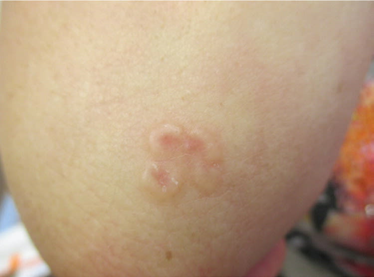
A 28-year-old woman presented with a fleshy, erythematous eruption with an annular configuration on her elbow that began 10 months earlier. The lesion was asymptomatic and had slowly increased in size. Her medical history was unremarkable, and she had no history of trauma.
Physical examination revealed erythematous, annular, nonscaly plaques with elevated and indurated borders (Figure 1). The plaques were composed of small flesh-colored papules and were localized to the right elbow.
Figure 1.
Question
Based on the patient's history and physical examination findings, which one of the following is the most likely diagnosis?
A. Granuloma annulare.
B. Nummular eczema.
C. Pityriasis rosea.
D. Plaque psoriasis.
E. Tinea corporis.
Discussion
The answer is A: granuloma annulare. Granuloma annulare is an idiopathic papular dermatosis. There are several subtypes of granuloma annulare, including localized, generalized, subcutaneous, arcuate, and perforating. This patient had the typical characteristics of localized granuloma annulare, with asymptomatic, firm, erythematous, violaceous, brown or flesh-colored, nonscaly papules. These plaques occur in an annular configuration and usually involve the extensor surfaces of distal extremities.1 As the condition progresses, some central involution occurs.
Lesions are often solitary or few in number. Sites of predilection include the lateral or dorsal surfaces of the hands and feet. Most cases occur before 30 years of age, and the condition is common in school-aged children.1,2 The female-to-male ratio is approximately 2:1.1,2 There have been reports of granuloma annulare after the use of medications such as gold, allopurinol, diclofenac, quinidine, calcitonin, amlodipine (Norvasc), daclizumab (not available in the United States), and angiotensin-converting enzyme inhibitors.1–3 In adults, the condition has been associated with type 1 diabetes mellitus, dyslipidemia, malignancy, and possibly autoimmune thyroiditis.2,4
Nummular eczema is characterized by well-demarcated, pruritic, coin-shaped eczematous patches that are scaly and sometimes lichenified. The lesions are symmetric and usually occur on the extensor surfaces of the hands, arms, and legs. The scales are thin and sparse.
Pityriasis rosea is characterized by an initial herald patch, followed by the development of a generalized, bilateral, symmetric, papulosquamous eruption. The long axes are along the skin lines of cleavage, known as Langer lines. The distribution on the back forms a Christmas tree pattern. Mild pruritus sometimes occurs.
Plaque psoriasis is characterized by sharply demarcated, erythematous, round or oval, usually pruritic plaques with loosely adherent silvery-white micaceous scales. Removal of the scales results in fine punctate bleeding (Auspitz sign). The lesions are usually distributed symmetrically on the knees, elbows, and scalp.
Tinea corporis presents as a well-demarcated, sharply circumscribed, annular, erythematous, mildly pruritic plaque with a raised leading edge and scaling. The lesion spreads centrifugally, leaving the characteristic central clearing commonly known as ringworm.
Summary Table
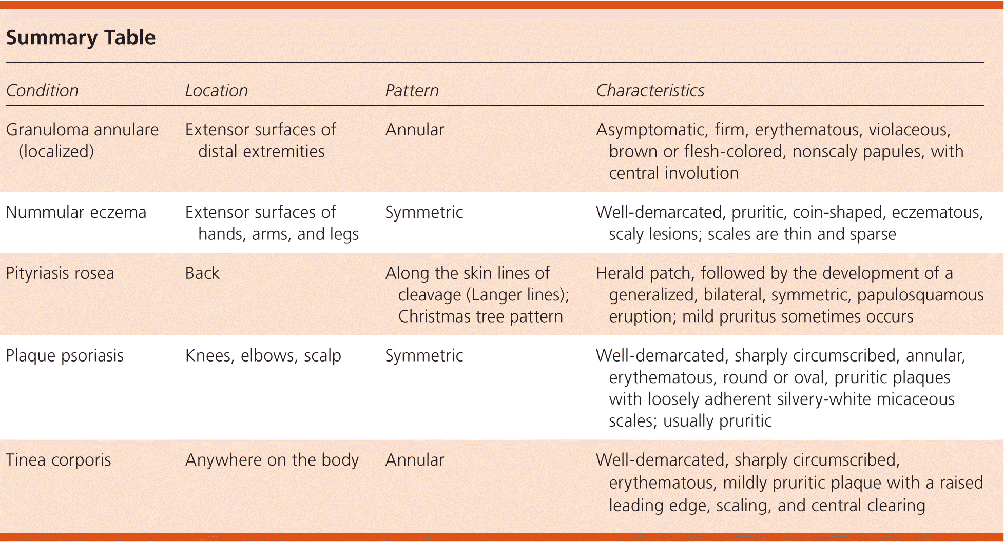
| Condition | Location | Pattern | Characteristics |
|---|---|---|---|
| Granuloma annulare (localized) | Extensor surfaces of distal extremities | Annular | Asymptomatic, firm, erythematous, violaceous, brown or flesh-colored, nonscaly papules, with central involution |
| Nummular eczema | Extensor surfaces of hands, arms, and legs | Symmetric | Well-demarcated, pruritic, coin-shaped, eczematous, scaly lesions; scales are thin and sparse |
| Pityriasis rosea | Back | Along the skin lines of cleavage (Langer lines); Christmas tree pattern | Herald patch, followed by the development of a generalized, bilateral, symmetric, papulosquamous eruption; mild pruritus sometimes occurs |
| Plaque psoriasis | Knees, elbows, scalp | Symmetric | Well-demarcated, sharply circumscribed, annular, erythematous, round or oval, pruritic plaques with loosely adherent silvery-white micaceous scales; usually pruritic |
| Tinea corporis | Anywhere on the body | Annular | Well-demarcated, sharply circumscribed, erythematous, mildly pruritic plaque with a raised leading edge, scaling, and central clearing |
