Hand-foot-and-mouth disease is caused by human enteroviruses and coxsackieviruses. Outbreaks can occur in the spring to fall and are common in North America, and most cases occur in patients younger than 10 years. Hand-foot-and-mouth disease is transmitted by fecal-oral, oral-oral, and respiratory droplet contact. Patients present with a low-grade fever, a maculopapular or papulovesicular rash on the hands and soles of the feet, and painful oral ulcerations. Lesions usually resolve in seven to 10 days; however, in rare cases, patients may have neurologic or cardiopulmonary complications. The differential diagnosis for childhood rashes and oral enanthems is broad and includes erythema multiforme, herpes, measles, and varicella. Treatment is supportive and directed toward hydration and pain relief as needed with acetaminophen or ibuprofen. Oral lidocaine is not recommended, and antiviral treatment is not available. The best methods to prevent the spread of hand-foot-and-mouth disease are handwashing and disinfecting potentially contaminated surfaces and fomites.
Hand-foot-and-mouth disease is a common viral disease that presents in primary care. This article presents a brief summary and review of the etiology, clinical features, diagnosis, prognosis, and evidence for the care of patients with hand-foot-and-mouth disease.
SORT: KEY RECOMMENDATIONS FOR PRACTICE
| Clinical recommendation | Evidence rating | Comments |
|---|---|---|
| The diagnosis of hand-foot-and-mouth disease should be based on presentation of a maculopapular or papulovesicular rash on the hands and soles of the feet and painful oral ulcerations.7 | C | Expert opinion from the Centers for Disease Control and Prevention |
| Supportive care should be used to treat hand-foot-and-mouth disease. Weight-based acetaminophen or ibuprofen may be used to treat fever and pain, but oral lidocaine is not recommended.7,39,40 | C | Consensus opinion (acetaminophen/ibuprofen); small randomized controlled trial and case report (lidocaine) |
| Handwashing decreases the risk of transmitting hand-foot-and-mouth disease.8,42 | C | Disease-oriented, retrospective studies |
A = consistent, good-quality patient-oriented evidence; B = inconsistent or limited-quality patient-oriented evidence; C = consensus, disease-oriented evidence, usual practice, expert opinion, or case series. For information about the SORT evidence rating system, go to https://www.aafp.org/afpsort.
Epidemiology
- Hand-foot-and-mouth disease was first described after an outbreak in Canada in the 1950s.1 It is caused by picornaviruses, specifically human enteroviruses and coxsackieviruses.2
- The most common viruses that cause hand-foot-and-mouth disease are enterovirus 71 and coxsackievirus A16.2 Currently, hand-foot-and-mouth disease is not listed as a notifiable condition in the United States by the Centers for Disease Control and Prevention; however, it has been a reportable illness in the Western Pacific region, where there are more severe outbreaks.3–5
- Coxsackievirus A6 can cause severe disease manifestations with atypical lesions such as vesicles, bullae, and scabs on the trunk, extremities, and face.6
- Spring to fall seasonal outbreaks of hand-foot-and-mouth disease are typical in North America and temperate zones.7,8 Years can pass between cyclical epidemics, during which time the pool of unexposed children increases.1
- Outbreaks of hand-foot-and-mouth disease are possible during the winter, and some are associated with coxsackievirus A6.2 Year-round outbreaks are common in tropical zones.8
- Most cases occur in patients younger than 10 years,1 and the largest incidence is within the first five years of life.9
- Health care professionals working with children are at risk of contracting hand-foot-and-mouth disease, and males and females are equally affected.2
- Hand-foot-and-mouth disease has a low fatality rate in uncomplicated cases in the United States (0.06% to 0.11%).10 However, there were 10.7 million cases in China between May 2008 and June 2014, with 3,046 deaths attributed to neurologic and cardiopulmonary complications.5 Patients with more severe disease are more likely to have been infected with enterovirus 71.5
Transmission
- Humans are the only carrier for hand-foot-and-mouth disease–causing viruses.1 The disease is spread by fecal-oral, oral-oral, and respiratory droplet contact.10
- The patient is most infectious during the first week of illness7; however, an active virus may be present in the stool for up to four to eight weeks.10 Therefore, the household transmission rate for hand-foot-and-mouth disease enterovirus 71 is 52% to 84%.10
- Incubation range is estimated to be three to six days.8
- Lack of access to clean water partially explains the burden of disease in the developing world and Asia, where hand-foot-and-mouth disease is a significant public health threat.2
Clinical Features
Hand-foot-and-mouth disease is a clinical diagnosis based on the presentation of a low-grade fever with a maculopapular or papulovesicular rash on the hands (Figure 111 ) and soles of the feet (Figure 211 ) and by painful oral ulcerations.7 If the diagnosis is unclear, serologic and polymerase chain reaction studies may be obtained to detect enterovirus or coxsackievirus.1,4,5,12
FIGURE 1.
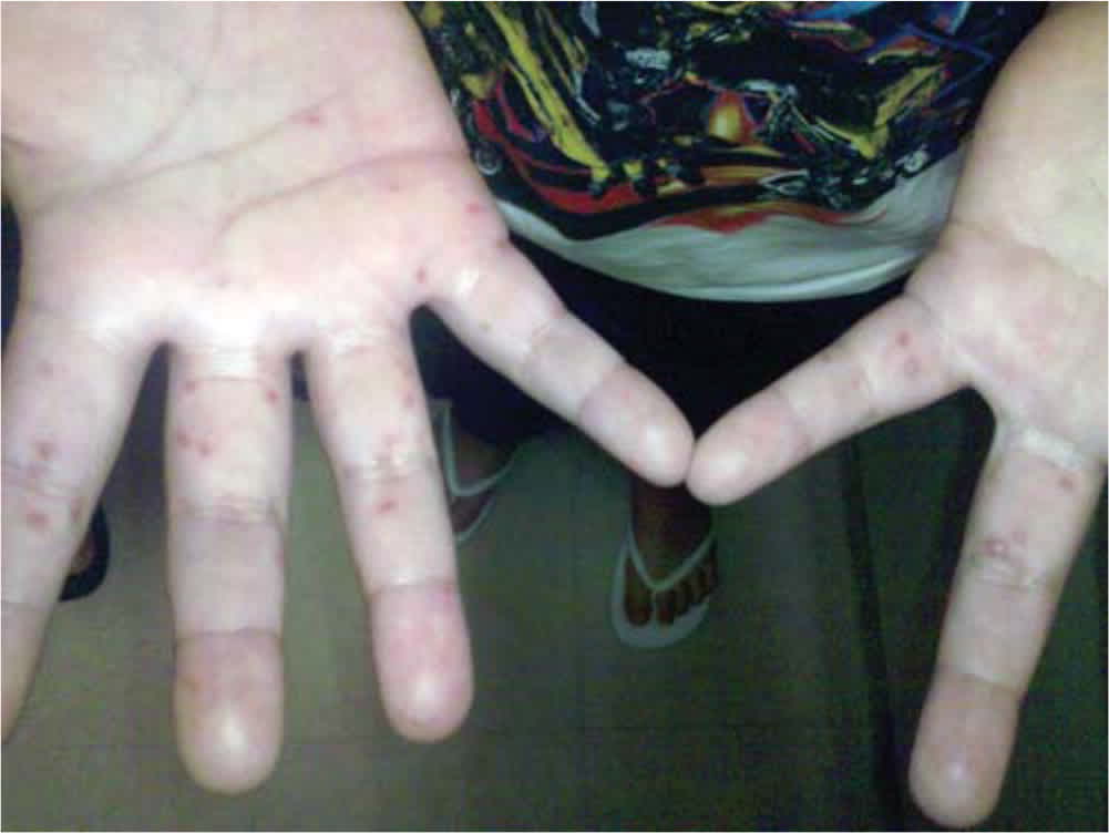
Maculopapular lesions on the palms of a patient with hand-foot-and-mouth disease.
Reprinted with permission from Pillai AS, Medina D. Rash in an eight-year-old boy. Am Fam Physician. 2012; 86(12):1141.
FIGURE 2.
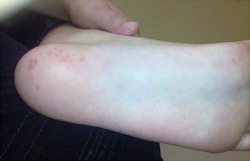
Maculopapular lesions on the soles of a patient with hand-foot-and-mouth disease.
Reprinted with permission from Pillai AS, Medina D. Rash in an eight-year-old boy. Am Fam Physician. 2012;86(12):1141.
- Skin lesions are typically 2 mm to 6 mm in diameter, have an erythematous halo, and evolve into vesicles that rupture and leave painless shallow ulcers that do not scar.4
- Oral enanthems of painful ulcerations typically affect the posterior oral cavity, including the soft palate. Lesions may also affect the tongue and buccal mucosa, and pain may cause dehydration4 (Figure 3).
- Lesions resolve in seven to 10 days.5
- Patients may have atypical skin lesions, including hemorrhagic or purpuric lesions; bullae and pustules; trunk, cheek, or genital involvement; palm and sole of the feet desquamation; and accentuation in areas of atopic dermatitis (eczema coxsackium).7,13
- The disease may be associated with delayed nail separation or horizontal nail ridges or grooves.1
- Rare neurologic complications can occur such as aseptic meningitis, acute flaccid paralysis, and encephalomyelitis, especially with enterovirus 71.5
- Other rare complications include pulmonary edema, pulmonary hemorrhage, and cardiorespiratory failure.4
FIGURE 3.
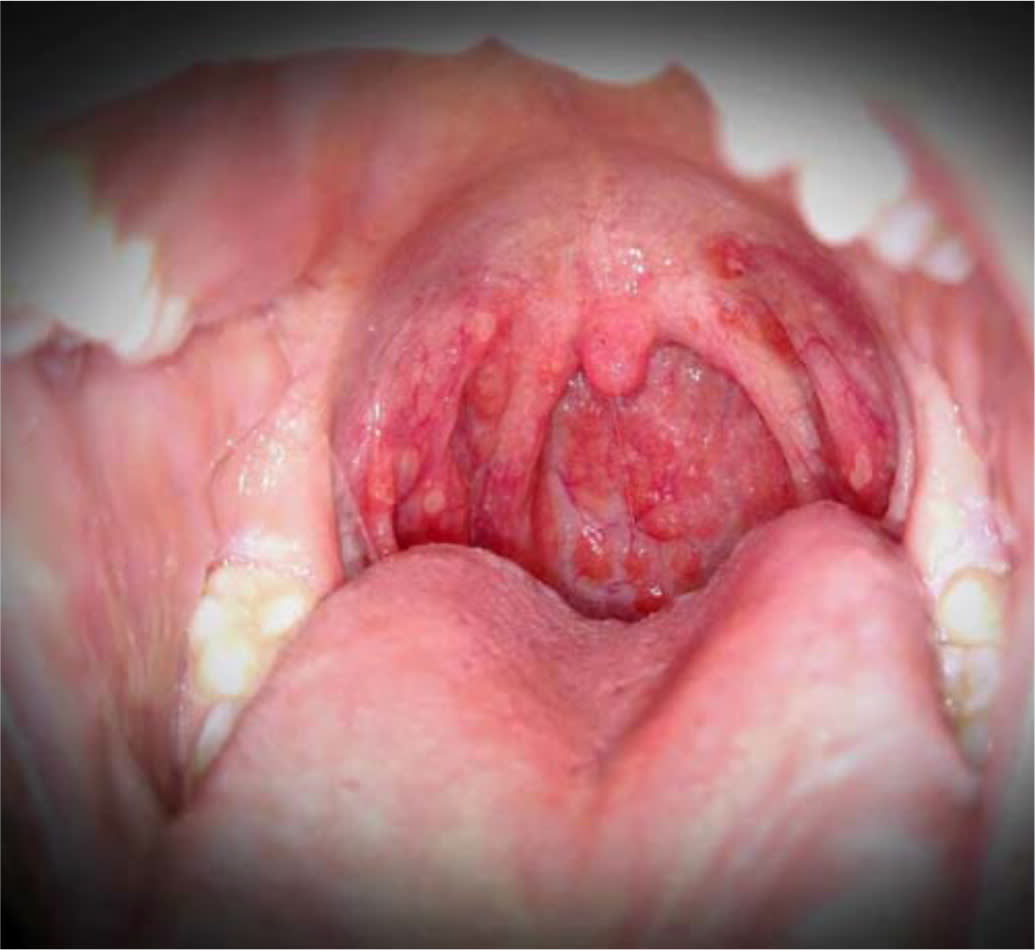
Oral ulcerations in a patient with hand-foot-and-mouth disease.
Differential Diagnosis
- Differential diagnosis includes diseases that feature maculopapular or papulovesicular rashes and/or oral lesions (Table 114–38 ).
- Aphthous ulcers and herpetic gingivostomatitis are typically limited to the oral cavity or surrounding skin.14,19
- Herpangina caused by the same agents as hand-foot-and-mouth disease is limited to the oral cavity without skin involvement.18
- Pemphigus vulgaris and Behçet syndrome include oral lesions and involve multiple systems. Both require recognition, further investigation, and treatment.17,22
- Herpes and varicella rashes have characteristic vesicles and erythema.30,38
- Atopic dermatitis is usually recurrent and has typical age-related distribution of lesions.24
- Scabies is intensely pruritic and associated with a linear distribution of lesions attributed to mite burrows.35
- Erythema multiforme major presents as target lesions on the face and limbs.27
- Bullous impetigo causes flaccid bullae that affect the trunk and extremities.26
- HIV should be considered with skin rash or oral lesions if risk factors are present.
TABLE 1. Differential Diagnosis of Hand-Foot-and-Mouth Disease
| Condition | Pathogenesis | Clinical presentation and diagnosis | Treatment | |||
|---|---|---|---|---|---|---|
| Oral enanthem | ||||||
| Aphthous ulcers | Unknown | Shallow, round, painful ulcers, measuring up to 1 cm, with surrounding erythema and pseudomembrane14 Simple aphthae resolve in one to two weeks, not associated with skin lesions Complex aphthae tend to be larger, occur more frequently, and may indicate systemic disease (e.g., gluten sensitive enteropathy), HIV, cyclic neutropenia, systemic lupus erythematosus, inflammatory bowel disease, periodic fever, aphthous stomatitis, pharyngitis, or cervical adenitis syndrome14 | Simple aphthae: supportive care Complex aphthae: treat underlying cause Pain relief: chlorhexidine (Peridex) mouthwash, lidocaine spray or ointment, anti-inflammatory or corticosteroid pastes or mouthwashes15,16 | |||
| Behçet syndrome | Unclear etiology, associations with human leukocyte antigen-B51 allele, postulated environmental triggers17 | Oral aphthae, genital ulcerations, or recurrent uveitis May have arthralgia, vascular or neurologic lesions Oral lesions are painful, round, with an erythematous border, and are 1 cm to 3 cm in diameter or larger17 | Corticosteroids, azathioprine (Imuran), cyclophosphamide, methotrexate, interferon alpha, ustekinumab (Stelara), infliximab (Remicade), etanercept (Enbrel), adalimumab (Humira)17 | |||
| Herpangina | Coxsackievirus, echovirus18 | Oral vesicles that form ulcers with associated inflammation Coxsackievirus A subtypes 1–6, 8, 10, and 2219 Thought to be on a continuum with hand-foot-and-mouth disease | Supportive care | |||
| Herpetic gingivostomatitis | Herpes simplex virus 1 and 2 | Fever, anorexia, lymphadenopathy, oral erythema and small, oral vesicles on the palate, tongue, gingiva, and oral mucosa that form ulcers that may become confluent; vesicles may be present on lips; Tzanck cells may be present, diagnosis can be made by culture or immunologic assay19,20 | Supportive care; acyclovir started in the first 72 hours resulted in faster resolution of oral lesions21 | |||
| Pemphigus vulgaris | Caused by desmosome autoantibodies22 | Oral mucosal bullae and erosions of lips, tongue, and oropharynx; may affect eyes and genital area; potentially life-threatening22 Diagnostic testing with direct immunofluorescence microscopy or serum testing | Corticosteroids, azathioprine, cyclophosphamide, intravenous immunoglobulin22 | |||
| Maculopapular or vesicular exanthem | ||||||
| Atopic dermatitis | Genetic, immunologic, and environmental factors23 | Erythematous plaques and vesicular lesions, excoriation, dry skin Younger children with lesions on extensor surfaces, cheeks; older children lesions on flexor surfaces; lesions on hands and feet common24 | Avoid triggers (e.g., cold weather, frequent hot baths, fragrances, detergents) Emollient creams, topical corticosteroids24; oral agents for severe cases25 | |||
| Bullous impetigo | Staphylococcus aureus | Superficial vesicles progress to flaccid bullae that rupture; collarette of scale surrounding blister at periphery of lesion; tends to affect trunk, extremities and moist, intertriginous areas; does not scar, systemic symptoms uncommon26 | Topical mupirocin (Bactroban) or retapamulin (Altabax); for more extensive disease or inability to tolerate topical therapy, may use amoxicillin/clavulanate (Augmentin), cephalexin (Keflex), dicloxacillin, doxycycline, or trimethoprim/sulfamethoxazole26 | |||
| Erythema multiforme | Immune mediated, often secondary to infection (specifically herpes simplex virus and Mycoplasma pneumoniae), may also be secondary to drugs and other causes | Trunk, limb, and face distribution, erythema multiforme minor limited to the skin, erythema multiforme major involves mucosal membranes; skin lesions < 3 cm in diameter; two concentric, colored rings surround dusky central zone; affects < 10% of body surface area, often elevated C-reactive protein level27 | Supportive care; if caused by a drug, discontinue that agent; if secondary to herpes simplex virus, consider antiviral therapy; corticosteroids may be used in severe cases, although controlled studies are lacking28 | |||
| Herpes | Herpes simplex virus 1 and 2 | Fever, pruritus,19 maculopapular and vesicular rash29,30; lesions may appear on areas in contact with oral herpes (e.g., herpetic whitlow), in areas prone to bodily contact (e.g., herpes gladiatorum), or on sites of previous atopy (e.g., eczema herpeticum31) | Acyclovir, famciclovir, or valacyclovir (Valtrex)30 | |||
| Measles | Measles virus | Respiratory spread; presents with fever, cough, coryza; Koplik spots (white papules) may present on buccal mucosa before maculopapular rash that starts on head and spreads distally Complications include pneumonia, keratoconjunctivitis, encephalomyelitis32 | Supportive treatment; vitamin A supplementation; measles may be prevented with routine childhood immunization; measles cause 100,000 deaths per year, worldwide32 | |||
| Rocky Mountain spotted fever | Rickettsia rickettsii, transmitted by infected tick (e.g., American dog tick, Rocky Mountain wood tick) | History of a tick bite (50% to 60% of patients), headaches, fever, fatigue, nausea, photophobia; rash starts with blanching, erythematous macules and papules on wrist and ankles, spreads centripetally; may ulcerate Complications include congestive heart failure, dysrhythmia, seizures, nerve palsies33 | Doxycycline; preventive measures include avoiding tick-infested habitats, tick repellant, full body skin examinations after exposure to areas with ticks33 | |||
| Scabies | Sarcoptes scabiei hominis34 | Linear distribution of papules corresponding with mite burrows; typical distribution includes hands, feet, skinfolds, genitalia; intense pruritus, worse at night; mites can be visualized in skin scrapings by microscope35 | Permethrin cream 5% (Elimite); wash all clothing, bedding, and towels in hot water; treat close contacts35 | |||
| Stevens-Johnson syndrome | Delayed-type hypersensitivity reaction usually associated with drugs | Fever, malaise prodrome; painful skin and mucous membrane (i.e., eye, mouth, and genital) lesions; erythematous skin with blister formation and flat atypical target lesions; pulmonary, renal, and hepatic involvement common; < 10% of skin surface area involved36 | Discontinue causative drug; refer to specialized units (e.g., burn centers); may consider corticosteroids, intravenous immunoglobulin, and/or cyclosporine A36 | |||
| Varicella (chickenpox) | Varicella zoster virus | Generalized, itchy, vesicular rash; fever, malaise; may cause pneumonitis, hepatitis, encephalitis, skin rash may become secondarily infected37; rash starts on face and trunk and spreads to rest of body; starts with macules and progresses to papules and vesicles; lesions visible in all stages at the same time as each other; symptoms last four to seven days38 | May use acyclovir within 24 hours of rash onset, or later in severe cases or in patients who are immunocompromised37; prevent with vaccination; avoid aspirin, may consider corticosteroids | |||
Treatment
Management is supportive and directed toward the relief of pain, lowering of fever, and adequate oral hydration because of the self-limiting nature of hand-foot-and-mouth disease.
- Discomfort because of pain or fever can be treated with weight-based acetaminophen or ibuprofen.7
- Oral application of topical lidocaine is not recommended for use in children because of the lack of benefit39 and the potential for harm.40
- Antiviral treatments are not available. One clinical trial of acyclovir (n = 13) reported a reduction of fever and skin changes within 24 hours; however, more evidence is needed.41
- Indications for hospitalization include a failure to maintain adequate hydration or the development of neurologic or cardiopulmonary complications.4
- Intravenous immunoglobulin is not recommended. In Asia, intravenous immunoglobulin is used in severe cases because of the potential benefit in stopping the progression to cardiopulmonary failure based on retrospective data; however, more prospective evidence is needed.4
Prevention
Handwashing stops the spread of hand-foot-and-mouth disease, specifically after diaper changes and toileting, and before eating.7,42,43
- In China, children who “always wash” hands before meals were less likely to contract the disease.8
- Disinfect surfaces and fomites (e.g., toys), avoiding close contact and the sharing of personal items such as utensils and cups with infected persons.7,43
- Breastfeeding does not impact the incidence of hand-foot-and-mouth disease. Mothers do not need to stop breastfeeding to prevent transmission of disease.8
- There are no vaccines or chemoprophylaxis agents available to prevent hand-foot-and-mouth disease and herpangina.7,44
- In the United States, exclusion from childcare does not reduce the spread of the disease and is not recommended unless the child is unable to participate or staff are unable to care for the child without compromising the care of other children.45
Data Sources: Sources consulted for this article include PubMed from the National Library of Medicine, Essential Evidence Plus, the Cochrane Database of Systematic Reviews, the Centers for Disease Control and Prevention, and the World Health Organization. Search terms included hand-foot-and-mouth disease, herpangina, and maculopapular exanthems. Search dates: October 2018, January 2019, and June 2019.
Editor's Note: Dr. Saguil is a contributing editor for AFP.
The views expressed in this article are the authors' own and do not necessarily reflect the views of the U.S. Army, U.S. Navy, U.S. Air Force, the Department of Defense, or the U.S. government.
