
Am Fam Physician. 2019;100(12):773-774
Author disclosure: No relevant financial affiliations.
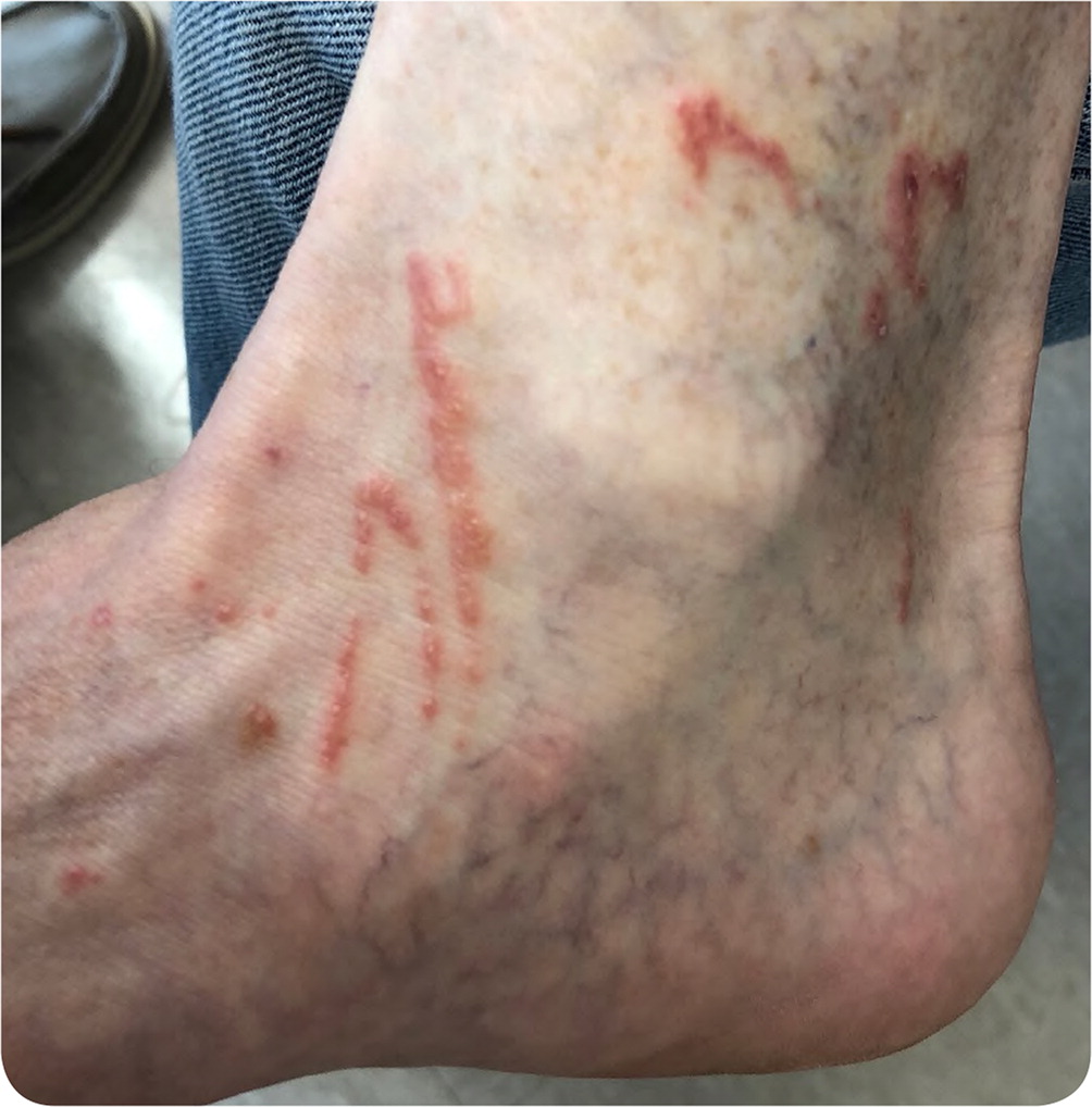
A 65-year-old man presented with a rash on his right ankle that developed four days prior. The rash began as a single papule but progressively spread. The patient reported itching but no pain or tenderness. He applied bacitracin and triamcinolone to the rash without improvement. The patient had a history of basal cell carcinoma, actinic keratosis, and seborrheic keratosis. He had recently visited the North Carolina coast immediately after a hurricane impacted the area. On physical examination, he was in no acute distress, and his vital signs were normal. A vesicular rash extended approximately 3 cm × 1 cm anterior and slightly superior to the right medial malleolus in a serpentine pattern. A second, smaller lesion was located directly superior to the same malleolus (Figure 1).
Question
Discussion
The most common causative organism is the hookworm larvae of a cat or dog (Ancylostoma braziliense or caninum). Larval development is promoted by warm, humid conditions leading to ground infestation. Transmission often occurs from walking barefoot on contaminated sand or soil. Natural disasters, such as floods and hurricanes, can lead to the additional spread of pathogens into the environment. Cutaneous larva migrans occurs in Southeast Asia, Central and South America, the Caribbean, and the southeastern coast of the United States. Diagnosis is based on clinical history of exposure and the characteristic serpiginous lesion.
Actinic keratosis is a precancerous cutaneous lesion that occurs in sun-exposed areas, such as the head, neck, forearms, and hands. It is most common in older people with fair skin. The rash is erythematous and scaly.
Impetigo is a skin infection caused by Staphylococcus aureus or Streptococcus pyogenes that is common in children. The rash usually presents on the face or upper extremities as a papule that progresses to a vesicle and then a honey-crusted lesion.
Myiasis is caused by infection from fly larvae and commonly occurs in tropical areas. It presents as a pruritic, painful, erythematous nodule with a central perforation.3 There is commonly pain and movement within the nodule.
Strongyloidiasis is an acute or chronic infection that may be secondary to a soil-transmitted parasitic nematode; human disease is primarily caused by Strongyloides stercoralis. It presents as a rash similar to cutaneous larva migrans but typically with gastrointestinal symptoms, usually diarrhea.4
| Condition | Description |
|---|---|
| Actinic keratosis | Precancerous lesion on sun-exposed areas, often in older adults with fair skin; lesions are erythematous and scaly |
| Cutaneous larva migrans | Erythematous papule on the lower extremity that evolves into the characteristic serpiginous lesion; caused by infection with hookworm larvae |
| Impetigo | Papule that progresses to a vesicle and then a honey-crusted lesion; commonly occurs on the face and upper extremities; skin infection caused by Staphylococcus aureus or Streptococcus pyogenes; common in children |
| Myiasis | Pruritic, painful, erythematous nodule with a central perforation; pain and movement within the nodule; caused by infection with fly larvae |
| Strongyloidiasis | Rash similar to cutaneous larva migrans but with gastrointestinal symptoms, usually diarrhea; caused by Strongyloides stercoralis infection |
