
Am Fam Physician. 2021;103(9):559-560
Author disclosure: No relevant financial affiliations.
A 69-year-old patient was referred to the wound care clinic from a rehabilitation facility for evaluation of gluteal skin ulcers. The patient had no fever or chills. The medical history included atrial fibrillation (without anticoagulation), uncontrolled type 2 diabetes mellitus, peripheral neuropathy, and stage 3 chronic kidney disease.
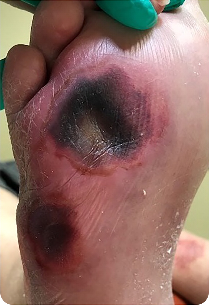
On physical examination, the patient had erosive dermatitis in moist gluteal areas. Further examination revealed nontender, discolored lesions on the plantar surface of the right foot (Figure 1).
Question
Based on the patient's history and physical examination findings, which one of the following is the most likely diagnosis?
A. Arterial embolization.
B. Calciphylaxis.
C. Deep tissue injury.
D. Morel-Lavallée lesions.
E. Morphea.
Discussion
The answer is C: deep tissue injury. Deep tissue injury presents as a localized area of dark purple or maroon intact skin or blood-filled blisters. It is caused by underlying soft tissue damage from pressure and/or a shearing force, and a likely combination of ischemic and reperfusion injury.1,2 Deep tissue injuries make up approximately 9% of all pressure injuries and are often poorly recognized by health care professionals.3 The lesions form rapidly, with 24 to 72 hours between soft tissue damage and onset of discoloration.4 Even with optimal treatment, the affected areas may progress to stage 3 and 4 pressure injury. Immediate off-loading, restoration of perfusion, improved patient nutrition, and diligent topical care are essential. The presence of both moisture-associated dermatitis and deep tissue injury may suggest neglect and could lead to medicolegal issues.
This patient likely developed deep tissue injuries as a result of pressure on the sole of the foot from contact with the railing when sliding down the bed. Arterial studies of the lower extremities did not reveal peripheral vascular disease, and radiographs did not show bone injuries. Immediate off-loading of the pressure was initiated. Despite aggressive therapy, necrosis developed down to the plantar fat pad. Necrotic tissue was debrided mechanically and enzymatically. The patient recovered completely after five months of treatment.
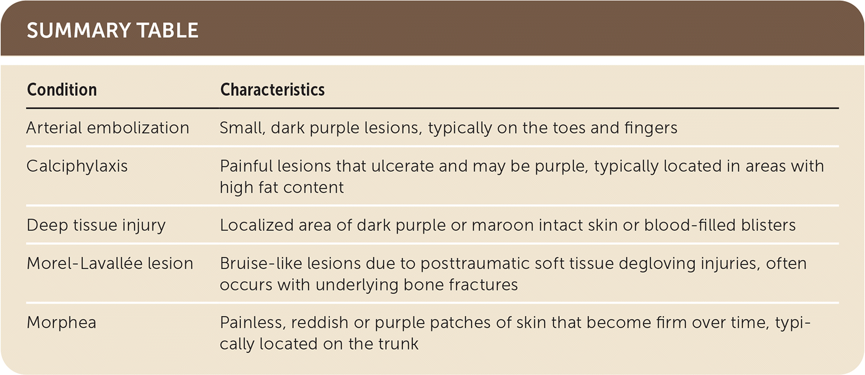
Arterial embolization often presents as small, dark purple lesions due to underlying necrosis of subcutaneous tissues. These lesions are painful and often located on the toes and fingers.
Calciphylaxis occurs in patients with chronic kidney disease and presents as painful lesions that ulcerate. The lesions may be purple because of underlying tissue necrosis and are usually located in areas with high fat content.
Morel-Lavallée lesions are bruise-like lesions due to posttraumatic soft tissue degloving injuries that cause blood and serosanguinous fluid to accumulate between skin or subcutaneous tissues and fascia. Morel-Lavallée lesions often occur with underlying bone fractures.5
Morphea is a form of scleroderma that presents as painless, reddish or purple patches of skin. The lesions become firm over time because of sclerosis and are typically located on the trunk.
| Condition | Characteristics |
|---|---|
| Arterial embolization | Small, dark purple lesions, typically on the toes and fingers |
| Calciphylaxis | Painful lesions that ulcerate and may be purple, typically located in areas with high fat content |
| Deep tissue injury | Localized area of dark purple or maroon intact skin or blood-filled blisters |
| Morel-Lavallée lesion | Bruise-like lesions due to posttraumatic soft tissue degloving injuries, often occurs with underlying bone fractures |
| Morphea | Painless, reddish or purple patches of skin that become firm over time, typically located on the trunk |
