Am Fam Physician. 2021;104(3):305-308
Author disclosure: No relevant financial affiliations.
Case Scenario
A 78-year-old patient in good health has hypertension that is well controlled with medication. One fall afternoon, the patient was raking leaves when they suddenly passed out. The patient had no dizziness or other symptoms before or after the event. Their partner saw them fall and rushed over; the patient woke up instantly, felt fine, stood up, and started walking. The patient did not hit their head and was taken to the hospital, where a computed tomography (CT) scan of the head, carotid Doppler, and echocardiography were performed before the patient was admitted to a telemetry unit. The patient's blood pressure was elevated at times, and carotid artery studies showed mild stenosis; therefore, the patient was started on atorvastatin (Lipitor), and their lisinopril dose was increased. No specific cause of syncope was found after two days of monitoring. The patient was scheduled for follow-up appointments with a neurologist and cardiologist. The day after discharge from the hospital, the patient passed out again. The patient's blood pressure was 90/60 mm Hg.
Clinical Commentary
BENEFITS AND RISKS OF HOSPITALIZATION
Sometime in their lives, 40% of adults will have syncope, and many of them will go to the hospital for a workup and be admitted.1 Approximately 1.5% of all emergency department visits are for syncope, and between 27% and 35% of these people are admitted to the hospital. The average length of stay is two days, and the average cost per admission in 2011 was $28,000.2 Many people admitted for syncope were previously admitted for the same diagnosis and had the same workup; one study estimated that the rate of recurrent syncope admissions is 25%.2 That same study showed that 42% of patients with syncope had no identifiable cause, and the most common causes when found were hypokalemia, atrial fibrillation, ventricular tachycardia, dehydration, and hyponatremia. The mortality rate for patients with primary syncope is 0.2% over one year.2
The 2017 American College of Cardiology/American Heart Association/Heart Rhythm Society syncope guidelines present a data-driven algorithm to initiate a workup for syncope and determine which people are at high risk.3 After a detailed history, physical examination, and electrocardiography, most people can be identified as low risk based on normal findings or as higher risk based on abnormal or worrisome findings.4 In the latter category, the guidelines suggest other testing and treatment that may be beneficial as determined by the specific abnormal findings.
An analysis of a large cohort of patients who presented with syncope between 2004 and 2012 found that most people hospitalized were older and had more comorbidities than people evaluated as outpatients, putting them at higher risk of a secondary cause of syncope. Monitoring was the primary reason for admission. People who were hospitalized had a far higher 30-day and one-year mortality rate than those not hospitalized, which was likely related to their underlying diseases. Most causes of death in this cohort were unrelated to syncope.5 Hospitalization did not increase the chances of finding a life-threatening arrhythmia or a cause of syncope that was imminently dangerous. People admitted to the hospital were more likely to have nonfatal arrhythmias and nonarrhythmic causes of syncope identified earlier, without any change in mortality.6
Although most hospital admissions for syncope do not lead to the identification of an immediately life-threatening cause, hospitalization leads to more adverse events. One study found that 7.4% of people with syncope experienced a serious adverse event within 30 days if they were hospitalized, and 3.2% if not hospitalized.7 In a cohort of people at low risk who were admitted for syncope, 15% experienced adverse events in the hospital, including delirium, transfusion errors, falls, hypoglycemia, and medication errors. A total of 32% of people admitted to the hospital had unrelated incidental findings, leading to more testing and specialist referrals. Overall, patients who were admitted had an average of 11 diagnostic tests during their admission.8
TESTING IN THE HOSPITAL
The most common tests in people hospitalized for syncope are CT scan of the head, echocardiography, carotid Doppler, and cardiac monitoring. In one study, 76% of patients had a head CT scan, 69.7% had echocardiography, and 33%, had carotid Doppler.9 These tests were found to increase the total cost of hospitalization and the length of stay. The total cost of hospitalization for syncope among Medicare recipients is $2.4 billion per year9; therefore, the question is whether this testing helps uncover causes of syncope that are life-threatening or cannot be discovered in the outpatient setting.
A head CT scan is the most widely ordered test for syncope, but it rarely uncovers a cause. According to a Choosing Wisely recommendation from the American College of Emergency Medicine, the risks of a head CT scan outweigh the benefits in most cases of syncope, and it should not be ordered routinely in the absence of a head injury or signs of a stroke.10 However, a large meta-analysis found that up to two-thirds of patients presenting with syncope had a CT scan of the head.11 The diagnostic yield of those scans was 1.1% in the hospital and 3.8% in the emergency department, with most abnormal findings occurring in people with neurologic signs, trauma, or old age or in those using anticoagulants.11
Echocardiography is performed on 39% to 91% of people who present with syncope.12 The diagnostic yield of echocardiography in someone with normal history, examination, and electrocardiogram findings approaches zero; the cost per abnormality found is $60,000 to $132,000, and many of these abnormalities are not related to the cause of syncope and are not life-threatening.12 In people with abnormal electrocardiogram or cardiac examination findings, 29% of echocardiogram findings are abnormal; however, many of these tests do not reveal the cause of syncope and have been abnormal in the past.12 In people without cardiac risk factors who have an abnormal echocardiogram result, most have preexisting abnormalities or abnormalities that are not considered causative of syncope.13
Carotid ultrasonography is also commonly performed, despite clear guidelines from Choosing Wisely and other groups to limit its use. The American Academy of Neurology recommends against performing imaging of the carotid arteries for simple syncope in patients without other neurologic symptoms.14 Carotid ultrasonography is performed in approximately one out of six patients enrolled in Medicare who present with simple syncope (i.e., syncope with a negative initial workup and no worrisome signs or symptoms) and no neurologic signs, at an estimated cost of $33 million to $49 million annually.14 Even when carotid ultrasonography shows an abnormality, it is rarely diagnostic for syncope and seldom leads to a change in treatment.14 Between 1% and 4% of syncope cases are attributable to neurologic causes, and in almost all cases, patients present with neurologic signs and symptoms.15 According to the American Academy of Neurology, carotid ultrasonography should be reserved for people with focal weakness, signs of stroke, or carotid bruits.14
One of the primary reasons for hospitalization due to syncope is continuous telemetry. Few studies have demonstrated any mortality benefit, and people hospitalized for syncope typically do not have better survival rates from arrhythmia than those not hospitalized. One study demonstrated that in-hospital telemetry found significant arrhythmias in people with syncope who were older than 86 years or who presented with pulmonary edema. Telemetry revealed the cause of syncope in 17.6% of these patients, and 14.6% were transferred to the coronary care unit because of atrioventricular block or extreme bradycardia. However, even in this high-risk cohort, no one died from the arrhythmia, and monitoring had no demonstrable survival benefit.16
The in-hospital workup of syncope does not improve prognosis and may lead to more adverse consequences than if the workup was performed as an outpatient. Many of the diagnostic tests ordered in the hospital add to cost and clinical burden without reducing the risk of death. There is no evidence that performing necessary tests in the hospital provides any benefit compared with performing the same tests out of the hospital. Risk stratification tools have been validated to predict who needs more thorough follow-up after a syncope event,17,18 but the data indicate that even people at high risk do not necessarily benefit from hospitalization.

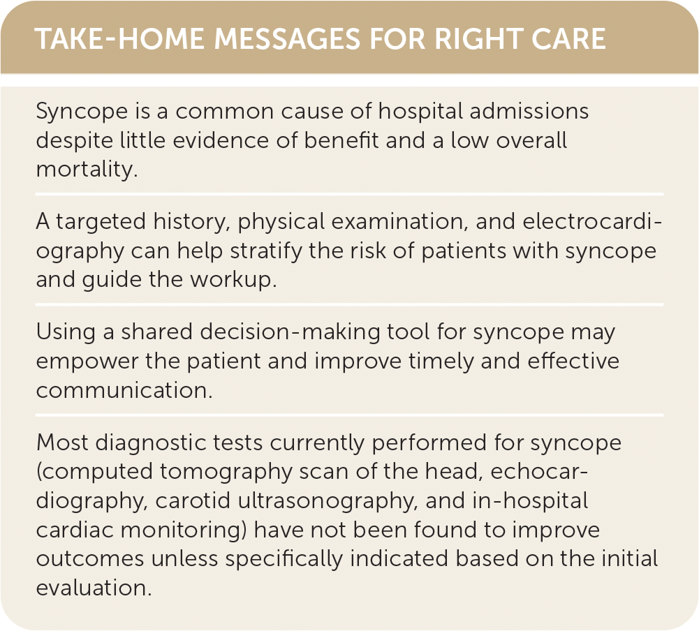
| Syncope is a common cause of hospital admissions despite little evidence of benefit and a low overall mortality. |
| A targeted history, physical examination, and electrocardiography can help stratify the risk of patients with syncope and guide the workup. |
| Using a shared decision-making tool for syncope may empower the patient and improve timely and effective communication. |
| Most diagnostic tests currently performed for syncope (computed tomography scan of the head, echocardiography, carotid ultrasonography, and in-hospital cardiac monitoring) have not been found to improve outcomes unless specifically indicated based on the initial evaluation. |
Patient Perspective
The dry recitation of the hospital's response to this patient's syncope reads like a primer for overuse. Our experience corroborates that extensive testing is a typical response to syncope, but it is largely ineffective and may cause harm. As patient advocates, we hear harrowing stories of overdiagnosis, misdiagnosis, and straight-out errors following syncope. One of the worst is very personal, involving a 19-year-old college student who experienced syncope while running in hot conditions.19 A blood test and electrocardiogram in the emergency department showed mild hypokalemia and a prolonged QT interval. During five days of evaluation in the hospital, the patient had more noninvasive tests, a cardiac catheterization, and an electro-physiology test with no clear findings. The patient's low potassium was never corrected, there was no warning to stop running, and the patient was given a clean bill of health. Three weeks after discharge, while running alone on the college campus, the student experienced a second syncope episode that was fatal.
Although the consequences were thankfully less dire, we have also experienced the two-day $28,000 workup for syncope—twice, with older family members who were later determined to have had known reactions to common drugs.
These examples illustrate two patterns in patient reports of their treatment after syncope. One is the underrated role of prescription drug adverse effects, a common and often poorly understood cause of syncope and presyncope symptoms. The primary care physician who knows the patient's history and medication list is in the best position to assess and adjust for possible medication-related effects. The other pattern is the misdiagnosis of long QT syndrome, which was due to potassium depletion in the college student, and greatly increases the risk of sudden cardiac death. Although it is relatively rare, long QT syndrome is often misdiagnosed. Our experience as patient advocates suggests that this may especially be the case in younger women, whose symptoms may sometimes be dismissed as psychiatric in origin.
Understanding that evaluation of syncope may be a complex undertaking, we support the emphasis on checking electrolyte levels and electrocardiogram results, especially in patients who experience syncope while exercising or working in hot conditions. Using a shared decision-making tool for syncope may empower the patient and improve timely and effective communication with the clinician or primary care physician.20
Resolution of Case
The patient followed up with their primary care physician, who reduced the lisinopril back to the previous dose and discontinued the atorvastatin due to lack of evidence of benefit.21 The patient's partner, who also came to the appointment, told the doctor that the patient had not been eating lunch lately and experienced more dizziness later in the day, which is the time when the first fainting episode occurred. The patient's presentation, history, examination, and electrocardiogram findings did not put them in a high-risk category; therefore, the patient was reassured and counseled to stay hydrated and to let the physician know if this happens again.
