A 16-year-old patient with no significant medical history presented with pain and new deformity of his left middle finger the Monday after playing in a Friday night football game. He reported that he jammed his finger while attempting to catch a punt return and dropped the ball. The athletic trainer evaluated the patient's finger on the sidelines and buddy taped it to the adjacent finger. He played the rest of the game without issue.
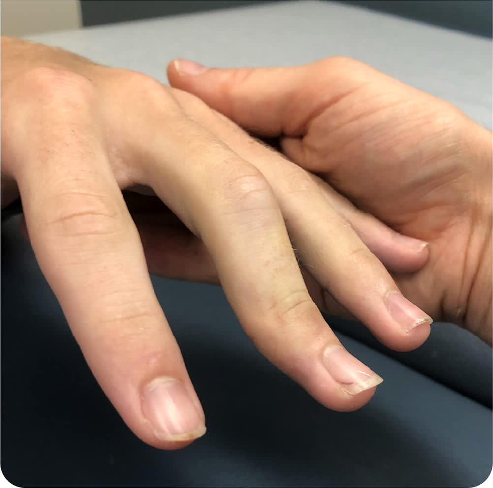
Over the weekend, the patient noticed that his injured finger was involuntarily flexed at the middle knuckle, and the tip was extended (Figure 1). He had no numbness or tingling. The middle knuckle was the most tender part of the injured finger, and the patient was apprehensive about moving it.
FIGURE 1
Question
Based on the patient's history and physical examination findings, which one of the following is the most likely diagnosis?
- A. Central slip injury.
- B. Jersey finger.
- C. Mallet finger.
- D. Sagittal band injury.
- E. Volar plate injury.
Discussion
The answer is A: central slip injury. The term jammed finger can refer to many injuries. The position of the finger during the injury, whether it is flexed or extended, and which joint is impacted are important factors in diagnosing a finger injury. The sport or activity associated with the injury can also be helpful. Radiography of the hand, particularly anteroposterior, lateral, and oblique views, should be performed to assess for dislocation, fracture, or level of tendon retraction and to guide management.1,2
A central slip injury typically occurs when an extended finger is forced into flexion at the proximal interphalangeal (PIP) joint. The extensor tendon is divided into three bands: the central slip and two lateral bands. The main function of the central slip is to extend the finger at the PIP joint. The lateral bands, which assist in flexion, are on the medial and lateral aspects of the proximal finger and terminate at the distal phalanx. A flexion deformity at the PIP joint can occur with complete rupture of the central slip. An extension deformity at the distal aspect of the injured finger (called a boutonnière deformity) can occur when the PIP joint buttonholes volarly along with the lateral bands, which causes extension at the distal interphalangeal (DIP) joint. A boutonnière deformity can develop anytime following a complete central slip injury but is not always present.
The Elson test is used to evaluate the integrity of the central slip. The patient hangs the injured finger off the edge of a table and attempts to extend the finger at the PIP joint. A passive DIP joint should appear taut, whereas complete rupture of the central slip results in joint laxity relative to the uninjured fingers.
Jersey finger is an injury to the flexor digitorum profundus tendon, which terminates at the DIP joint and is responsible for flexion. The injury is caused by forced extension of a partially flexed finger, often from catching or jamming a finger on an opponent or a ball. The ring finger is commonly involved. Pain and swelling at the DIP joint may occur, with the inability to flex the finger.
Mallet finger refers to a drooping or flexed posture at the DIP joint of the injured finger. It commonly occurs during sports such as racquetball and basketball due to a sudden blow to the tip of a finger while in extension. This injury can range from extensor tendon damage to complete rupture or avulsion fracture. The mallet finger deformity is a result of unopposed activity of the flexor digitorum profundus.
The sagittal bands are ribbon-like ligaments that encircle the metacarpophalangeal joint to stabilize and centralize the extensor tendons during motion. Injury to the hand, such as during a boxing match, can result in partial or complete rupture of a sagittal band. Patients with this injury often report a popping or snapping sensation, with immediate pain and edema at the knuckle. Full extension of the finger may be difficult or impossible.3 When making a fist, the defect can be seen and felt as the extensor tendon slides to one side of the knuckle.
The volar plate is a multilayered condensation of fibrocartilaginous tissue between the flexor tendons and the palmar PIP joint capsule. Sudden forced hyperextension of the PIP joint and occasionally crush injuries can result in partial or complete volar plate and collateral rupture.4 With this injury, the PIP joint is usually swollen with bruising and tenderness on the volar aspect.
SUMMARY TABLE
| Condition | Characteristics | Treatment |
|---|---|---|
| Central slip injury | Forced flexion of an extended finger at the PIP joint; forced flexion deformity present at the PIP joint; forced extension deformity may be present at the DIP joint (boutonnière deformity) | Splint PIP joint in extension, with the DIP and metacarpophalangeal joints freely mobile, for six weeks followed by night splinting for six weeks |
| Jersey finger | Forced extension of a partially flexed finger; commonly involves the ring finger and ball sports; injury to the flexor digitorum profundus tendon with pain and swelling at the DIP joint and the inability to flex the finger | Baseline plain radiography and prompt surgical referral |
| Mallet finger injury | Sudden blow to the tip of an extended finger; drooping or flexed posture at the DIP joint | Continuous splinting in slight hyper-extension for eight weeks; flexion at the DIP joint should be strictly avoided during splinting |
| Sagittal band injury | Injury to the hand, such as during a boxing match, can result in partial or complete rupture of a sagittal band; popping or snapping with movement of extensor tendons | Splint in extension for four to six weeks; surgical referral required for chronic symptoms (> six weeks) |
| Volar plate injury | Sudden forced hyperextension at the PIP joint and occasionally crush injuries can result in partial or complete rupture; swelling of the PIP joint with bruising and tenderness on the volar aspect | Splint in partial flexion followed by finger motion exercises and hand therapy; surgical referral required for joint instability or a significant fracture |
DIP = distal interphalangeal; PIP = proximal interphalangeal.
