An 84-year-old man presented with a two-month history of bilateral breast enlargement. Left nipple discharge began 2 1/2 weeks before presentation. He had no discharge from his right nipple. The milky, beige discharge occurred daily, most notably in the early morning and evening, and sometimes contained bright red blood. It was nonpurulent. The patient reported intermittent tenderness in his breasts but had no other symptoms, including fever, chills, night sweats, unexpected weight loss, fatigue, or change in libido or sexual function. He did not have a history of similar symptoms, steroid use, or hormone therapy. The patient's medical history included atrial fibrillation, anticoagulation, hypertension, chronic heart failure, and arthritis treated with long-term opiate use.
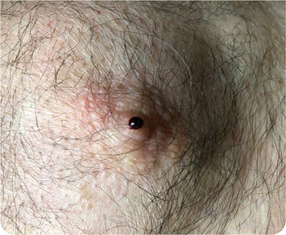
Physical examination revealed bilateral gynecomastia that was more pronounced on the left side. A 1-cm mass was palpated at the 10-o'clock position, about 2 to 4 cm from the left nipple. Another 1-cm mass was palpated at the 4-o'clock position, about 1 cm from the same nipple. One drop of bright red blood was expressed from the left nipple (Figure 1). No other masses were detected in either breast, and no axillary lymphadenopathy was palpated.
FIGURE 1
Question
Based on the patient's history and physical examination findings, which one of the following is the most likely diagnosis?
- A. Atypical ductal hyperplasia.
- B. Ductal carcinoma in situ.
- C. Fibrocystic breast changes.
- D. Intraductal papilloma.
- E. Unilateral mastitis.
Discussion
The answer is B: ductal carcinoma in situ (DCIS), which is uncontrolled growth of cells that line the breast ducts without spread through the walls of the ducts into surrounding breast tissue.1 DCIS accounts for approximately 20% of new breast cancer diagnoses. It is less common in men, representing an estimated 542 of the 2,710 new cases of breast cancer diagnosed in the United States in 2022.1
DCIS is usually asymptomatic and diagnosed by mammography, although unilateral nipple discharge occurs in some cases. Nipple discharge has a stronger association with breast cancer in men than women. Breast cancer should be highly suspected if nipple discharge is unilateral, bloody, serous, clear, spontaneous, or associated with a mass.2 Mammography and subareolar ultrasonography are usually diagnostic; however, patients with suspicious clinical findings should be referred to a surgeon for ductal excision, even with normal imaging findings. Cytology of the nipple discharge is not recommended because the absence of malignant cells does not exclude cancer.3 DCIS is treatable and has a low likelihood of metastasis if detected early. Late diagnosis is associated with progression to invasive ductal carcinoma.4
Atypical ductal hyperplasia is a premalignant growth of the cells that line the breast ducts. The condition is usually asymptomatic and detected incidentally on screening mammography. Atypical ductal hyperplasia increases the risk of breast cancer approximately four- or fivefold compared with the general population and is classified as a high-risk lesion.5 Atypical ductal hyperplasia is diagnosed by biopsy and treated with surgical excision.
Fibrocystic breast changes are the most common type of breast lesion in women but are uncommon in men. These benign nodular or glandular changes in breast tissue are typically asymptomatic but can feel firm on palpation and are sometimes associated with generalized breast pain, upper outer quadrant tenderness, or spontaneous nonbloody nipple discharge.6 Fibrocystic breast changes are thought to be caused by variations in estrogen levels, which fluctuate with the menstrual cycle.7
An intraductal papilloma is a benign fibrovascular tissue tumor within the milk ducts. It is a common cause of unilateral nonbloody nipple discharge but may present as a palpable mass around the nipple.8 A solitary intraductal papilloma forms in larger milk ducts and is often associated with nipple discharge. Having multiple papillomas slightly increases the risk of breast cancer.
Mastitis is caused by infectious or noninfectious breast inflammation. It may be associated with lactation. Nonlactational causes of mastitis include duct ectasia, idiopathic granulomatous inflammation, and foreign body reaction.9 Idiopathic granulomatous inflammation is most often mistaken for breast malignancy, presenting as nipple retraction, skin thickening, axillary adenopathy, ulceration, and abscess formation.
SUMMARY TABLE
| Condition | Characteristics |
|---|---|
| Atypical ductal hyperplasia | Premalignant growth of cells that line the breast ducts; usually asymptomatic and detected incidentally on screening mammography |
| Ductal carcinoma in situ | Uncontrolled growth of the cells that line the breast ducts without spread through the walls of the ducts into surrounding breast tissue; unilateral nipple discharge occurs in some cases |
| Fibrocystic breast changes | Benign nodular or glandular changes in breast tissue; typically asymptomatic; firm on palpation and sometimes associated with generalized breast pain, upper outer quadrant tenderness, or spontaneous nonbloody nipple discharge |
| Intraductal papilloma | Benign fibrovascular tissue tumors within the milk ducts; common cause of unilateral nonbloody nipple discharge; may present as a palpable mass around the nipple |
| Mastitis | Caused by infectious or noninfectious breast inflammation; presents as nipple retraction, skin thickening, axillary adenopathy, ulceration, and abscess formation |
