A six-month-old boy presented with recently developed vesiculopustular lesions on the soles of both feet. He was born at term with no perinatal issues. Two weeks earlier, he was treated with a topical corticosteroid for allergic contact dermatitis, which did not lead to marked improvement.
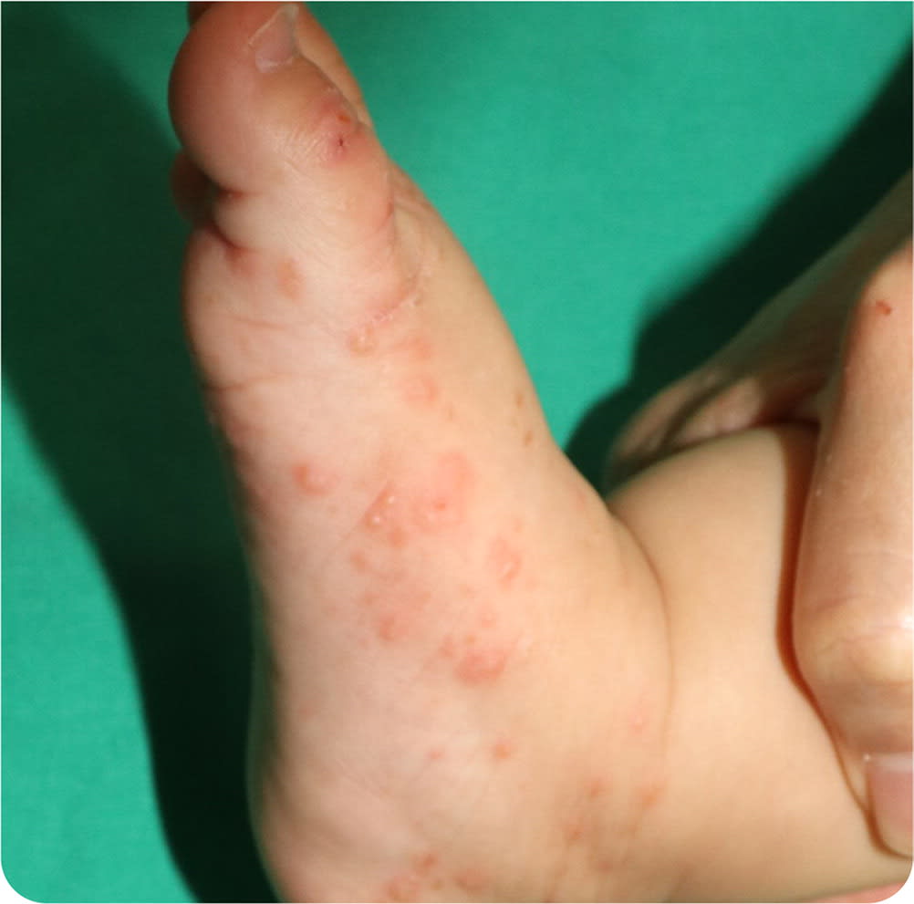
Physical examination revealed tiny, scaly vesicles and pustules on the soles extending to the lateral sides of the feet (Figure 1 and Figure 2). Some were erythematous, and others were the same color as the patient’s skin. Nodules were also found on the lower abdomen and perineal area. A few of the infant’s family members reported experiencing pruritus but did not have skin lesions.
FIGURE 1
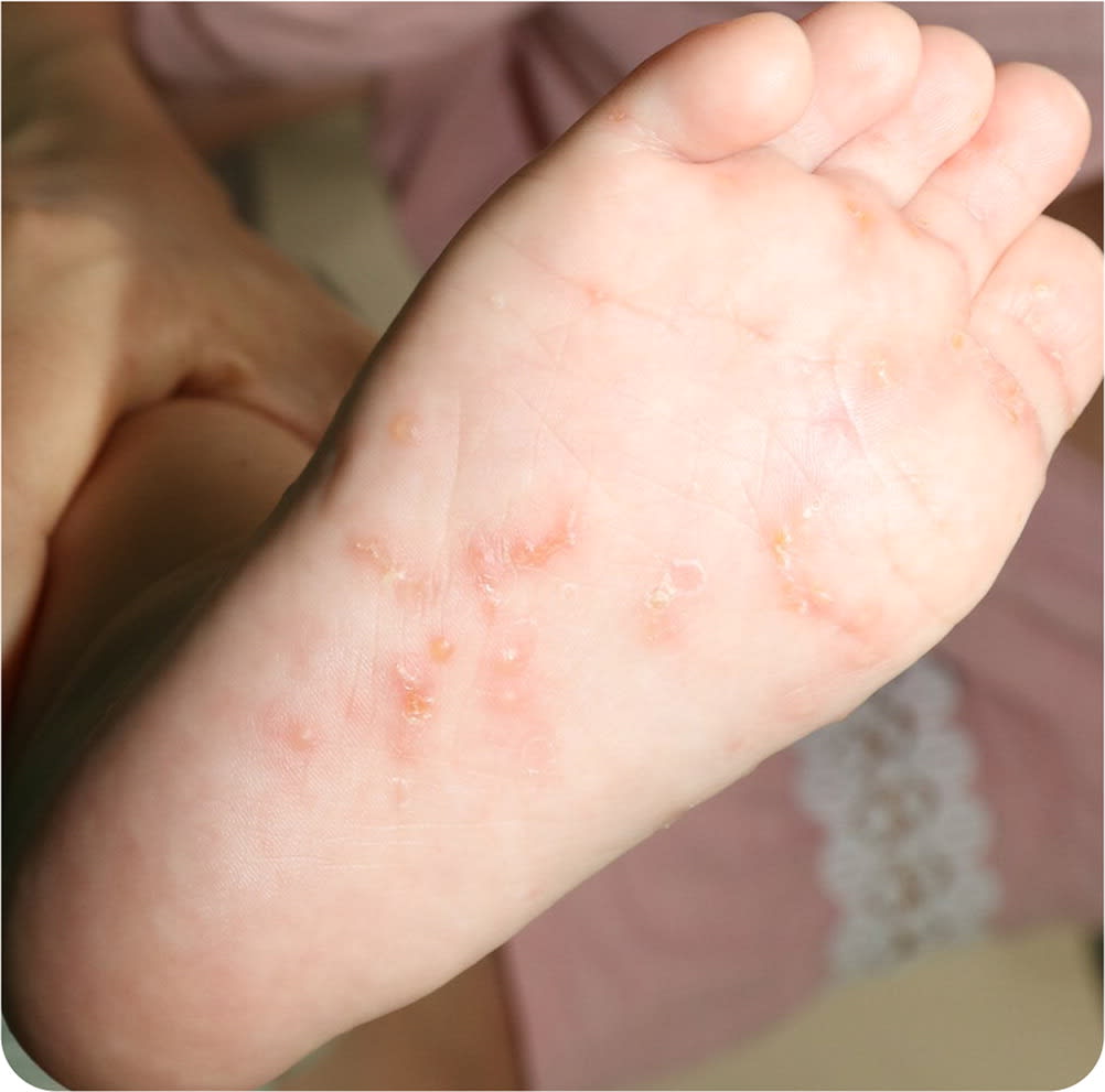
FIGURE 2
Question
Based on the patient’s history and physical examination findings, which one of the following is the most likely diagnosis?
- A. Bullous impetigo.
- B. Infantile acropustulosis.
- C. Infantile scabies.
- D. Langerhans cell histiocytosis.
Discussion
The answer is C: infantile scabies. Microscopic examination of skin scrapings revealed live mites. Infantile scabies was diagnosed, and the patient was treated with permethrin 5% cream. Oral antihistamines were also prescribed to alleviate pruritus. The lesions resolved without recurrent skin eruptions.
Diagnosis of infantile scabies can be challenging because the morphology and distribution of the skin lesions differ from those occurring in adults. Scabies infestation in adults is characterized by prominent nocturnal pruritus with excoriated papules and eczematous dermatitis, mainly on the interdigital webs, volar aspects of the wrists, axillae, external genitalia, and areolar tissue.
In contrast, scabies infestation in infants and young children is often associated with a pruritic papulopustular eruption. Vesicles, bullae, and pustules with erosions and ulcerations are much more common than papules. Lesions are often found on the face, scalp, palms, and soles. Additionally, pruritus and burrows are not as common as in adults.1 Once suspected, scabies is relatively easy to diagnose using skin scrapings. Early investigation is crucial for diagnosis and treatment.
Impetigo is a bacterial skin infection common in children. It is caused by Staphylococcus aureus and Streptococcus pyogenes. Impetigo shows two clinical patterns: bullous and nonbullous. Honey-colored, crusted erosions are characteristic of nonbullous impetigo. Bullous impetigo is caused by the exfoliative toxins of S. aureus and mostly occurs in newborns and infants. It also causes larger, thin-roofed blisters that arise on grossly normal skin areas, in addition to pustules and vesicles that rupture easily. The condition most commonly affects the face, but it can occur anywhere.
Infantile acropustulosis is an uncommon condition characterized by recurrent crops of tiny, intensely pruritic acral vesicles and pustules in young children. It is often found on the palms, soles, and sides of the feet. The intensity and duration of the eruptions generally decrease with each episode, and the condition spontaneously resolves in the first two to three years of life. Although several treatment options have been used, treatment is not usually necessary due to the benign course.2,3 Infantile acropustulosis is often misdiagnosed as scabies. Cases of infantile acropustulosis following scabies have been reported; however, any relationship between the two is unclear.4
Langerhans cell histiocytosis is a rare neoplastic disorder of myeloid dendritic cells. Although the condition can occur at any age, it is most common in young children. Clinical manifestations vary and can involve multiple organs, including organs of the bones, liver, spleen, and nervous system. Bones and lymph nodes are the most common sites, and the skin is affected in about 40% to 50% of patients. Identification of skin lesions is key for early diagnosis. Cutaneous manifestations typically present as rose-yellow, crusted papules or papulovesicles on the trunk, intertriginous areas, and scalp. It can also present as eczematous dermatitis, mimicking candidal intertrigo or seborrheic dermatitis.5
SUMMARY TABLE
| Condition | Characteristics |
|---|---|
| Bullous impetigo | Thin-roofed blisters; pustules and vesicles that rupture easily; most commonly affects the face, but it can occur anywhere |
| Infantile acropustulosis | Recurrent crops of tiny, intensely pruritic acral vesicles and pustules; often found on the palms, soles, and sides of the feet |
| Infantile scabies | Vesicles, bullae, and pustules with erosions and ulcerations are much more common in infants than papules and burrows; typically affects the face, scalp, palms, and soles |
| Langerhans cell histiocytosis | Rose-yellow crusted papules or papulovesicles on the trunk, intertriginous areas, and scalp; may involve multiple organs, including bones, liver, spleen, and nervous system |
