Am Fam Physician. 2009;79(11):963-970
A more recent article on vision loss in older adults is available.
Patient information: See related handout on glaucoma.
Author disclosure: Nothing to disclose.
Family physicians have an essential role in assessing, identifying, treating, and preventing or delaying vision loss in the aging population. Approximately one in 28 U.S. adults older than 40 years is visually impaired. Vision loss is associated with depression, social isolation, falls, and medication errors, and it can cause disturbing hallucinations. Adults older than 65 years should be screened for vision problems every one to two years, with attention to specific disorders, such as diabetic retinopathy, refractive error, cataracts, glaucoma, and age-related macular degeneration. Vision-related adverse effects of commonly used medications, such as amiodarone or phosphodiesterase inhibitors, should be considered when evaluating vision problems. Prompt recognition and management of sudden vision loss can be vision saving, as can treatment of diabetic retinopathy, refractive error, cataracts, glaucoma, and age-related macular degeneration. Aggressive medical management of diabetes, hypertension, and hyperlipidemia; encouraging smoking cessation; reducing ultraviolet light exposure; and appropriate response to medication adverse effects can preserve and protect vision in many older persons. Anti-oxidant and mineral supplements do not prevent age-related macular degeneration, but may play a role in slowing progression in those with advanced disease.
Loss of vision is one of the most feared complications of aging.1 Approximately one in 28 U.S. adults older than 40 years is visually impaired. Adults older than 80 years comprise 8 percent of the U.S. population, but account for 70 percent of cases of severe visual impairment.2 With a rapidly aging population, the number of persons with visual impairment in the United States and worldwide is expected to increase significantly in the next decade.
| Clinical recommendation | Evidence rating | References |
|---|---|---|
| All persons older than 65 years should be screened periodically for vision problems. | C | 10–14 |
| All older persons with diabetes should have a dilated eye examination within one year of diabetes diagnosis, and at least annually thereafter. | C | 17 |
| Tight control of glucose and blood pressure lowers the risk of progressive diabetic retinopathy. | A | 25, 31, 33–35 |
| Controlling blood pressure in older persons with and without diabetes may reduce the risk of ischemic vascular complications that can cause vision loss. | B | 36 |
| Smoking is linked to several causes of progressive visual impairment; smoking cessation counseling should be a routine aspect of care for older persons. | B | 38–40 |
| Antioxidant and zinc supplements, alone or in combination, do not prevent or delay onset of age-related macular degeneration. | A | 42 |
| Antioxidant and zinc supplementation may delay the progression of age-related macular degeneration in some persons with advanced disease. | B | 43 |
Adults with low vision are at high risk of depression, social withdrawal, and isolation.1,3 Vision loss in older persons is an independent risk factor for falls.4 Progressive vision loss can be associated with a syndrome of hallucinations which, although benign, can be disturbing to patients.5 Older persons with visual impairments are more likely to be institutionalized.6,7 Adults who are visually impaired are at risk of self-administered medication errors, which may result in an increased rate of hospitalization.8 A retrospective study of adult Medicare beneficiaries found that those with impaired vision incur significantly higher treatment costs for eye- and non–eye-related disease.9 Family physicians have an essential role in assessing, identifying, treating, and preventing or delaying vision loss in the aging population. Table 1 lists resources for older persons who are visually impaired.
| Lighthouse International |
| Telephone: (800) 829-0500 |
| Web site: http://www.lighthouse.org |
| Mission is to fight vision loss through prevention, treatment, and empowerment; provides free literature on glaucoma, macular degeneration, diabetes, cataracts, and other causes of impaired vision; Web site includes a searchable library, information on low vision rehabilitation services, and links to rehabilitation agencies, state agencies, advocacy groups, and optometrists and ophthalmologists nationwide |
| National Federation of the Blind |
| Telephone: (410) 659-9314 |
| Web site: http://www.nfb.org |
| More than 50,000 members and affiliates in all 50 states; the largest organization of persons in the United States who are blind; offers advocacy for persons with visual impairments; provides individual, group, and community education; supports research, technology, and programs encouraging independence and self-confidence |
| VisionAWARE |
| Telephone: (914) 528-5120 |
| Web site: http://www.visionaware.org/ |
| Primary focus is a “self-help for vision loss” Web site; produces self-help publications that provide adaptive techniques for persons with impaired vision; sponsors online education; committed to increasing the visibility of other organizations and resources that address the unmet needs of persons who are blind or who have low vision |
Screening for Vision Loss
| American Academy of Family Physicians10 |
| Older adults should be screened with the Snellen chart; interval not stated. |
| American Academy of Ophthalmology11 |
| Adults older than 65 years with no risk factors or identifiable eye disease should have a comprehensive medical eye evaluation every one to two years. |
| Canadian Task Force on the Periodic Health Examination12 |
| Visual acuity screening by chart should be part of the periodic health examination in adults older than 65 years. |
| Institute for Clinical Systems Improvement13 |
| Routine visual acuity screening should be performed in adults older than 65 years; interval not stated. |
| U.S. Preventive Services Task Force14 |
| Visual acuity screening by Snellen chart should be part of the periodic health examination in adults older than 65 years. |
Visual acuity screening is easily accomplished using a wall or handheld chart, such as the Snellen chart. The Early Treatment of Diabetic Retinopathy Study (ETDRS) chart (ftp://ftp.nei.nih.gov/charts/EC02_300.tif) was developed by the National Eye Institute and is the accepted international reference standard for determining visual acuity in clinical trials.15 Modifications of the ETDRS chart are available for use with low-literacy and non–English-speaking populations.
Visual acuity screening alone has limitations. Chart screening may not detect visual field loss or discriminate central versus peripheral vision loss, and is not sensitive to loss of accommodation, color vision change, or low-light vision impairment. Visual acuity screening alone cannot identify functional impairment or functional adaptation to vision loss.15 Inquiring about specific symptoms and the functional impact of vision loss should therefore be a part of the vision screening process.3,15
DISEASE-SPECIFIC SCREENING
Diabetic retinopathy is one of the leading causes of blindness in persons older than 40 years in North America.2 Diabetic retinopathy is classified as nonproliferative or proliferative, with or without macular edema. Up to 10 percent of persons newly diagnosed with diabetes will have retinopathy within one year of diagnosis. In persons with severe nonproliferative diabetic retinopathy, the risk of progression to vision-threatening proliferative retinopathy within one year is 50 to 75 percent.1 Recommendations for eye evaluation in persons with diabetes are summarized in Table 3.16,17
| Disease or condition | Recommended screening protocol |
|---|---|
| Age-related macular degeneration | No established screening protocol |
| Cataracts | No established screening protocol |
| Glaucoma | Insufficient evidence to recommend for or against routine screening in the general population; consider periodic screening with tonometry and automated visual field testing in high-risk groups (e.g., black persons, persons with strong family history)16 |
| Persons with diabetes and without retinopathy or with minimal nonproliferative retinopathy | Dilated eye examination within one year of diabetes diagnosis, and annually thereafter17 |
| Persons with diabetes and stable nonproliferative retinopathy | Dilated eye examination every six to 12 months17 |
| Persons with diabetes and unstable proliferative retinopathy or macular edema | Dilated eye examination every two to four months, depending on degree of disease and visual impairment17 |
Refractive error affects up to one third of persons older than 50 years.18 Refractive error can be detected with office-based visual acuity testing using the Snellen or ETDRS chart. Improvement in acuity with pinhole testing usually indicates some degree of potentially correctable refractive error. Older adults with refractive error should be referred to an eye care specialist for further evaluation and treatment.
Cataracts are the leading cause of blindness worldwide, and are the most common cause of low vision (but not blindness) in the United States.2 Cataracts can be readily detected with a handheld ophthalmoscope during vision screening. Cataracts that do not cause significant visual impairment may be followed medically. If the cataract is suspected of causing impaired vision, referral to an ophthalmologist is warranted.
The natural history and progression of open-angle glaucoma is poorly understood. Forty percent of persons with vision-threatening primary open-angle glaucoma have normal intraocular pressures, and the glaucoma will be missed by intraocular pressure measurements alone.16 Complete screening for open-angle glaucoma should include pressure measurement and automated visual field testing. The U.S. Preventive Services Task Force does not recommend for or against routine screening for glaucoma in older adults, but recognizes that some subgroups at higher risk (e.g., black persons) may benefit from periodic screening.16
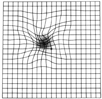
Age-related macular degeneration (AMD) is responsible for nearly 60 percent of blindness in adults of European descent older than 65 years.2 AMD is classified aswet(neovascularorexudative)ordry(non-neovascular or nonexudative). The Amsler grid may be used as a screening test for AMD. The grid detects linear distortion, metamorphopsia, and central scotomas, which are characteristic of AMD. The patient is instructed to look at the grid and report any wavy lines or areas that are missing or distorted (Figure 1).19 The specificity and sensitivity of the Amsler grid for detecting AMD in primary care practices are not known.

SCREENING FOR MEDICATION ADVERSE EFFECTS
| Drug or product | Effect (certain) | Effect (probable or possible) | Action |
|---|---|---|---|
| Amiodarone (Cordarone) | Blepharoconjunctivitis, colored halos around lights, glare, hazy vision, thyroid ophthalmopathy | Corneal ulceration, loss of eyelashes or eyebrows, nonarteritic ischemic optic neuropathy, pseudotumor cerebri | Consider eye examination every six to 12 months (for lifetime) because of the long half-life of the drug (controversial); if symptomatic, consider other causes of nonarteritic ischemic optic neuropathy and refer to an eye care specialist |
| Bisphosphonates (except risedronate [Actonel])* | Painful ocular inflammation including episcleritis, nonspecific conjunctivitis, scleritis | Diplopia, eyelid edema, glaucoma (with alendronate [Fosamax] only) | Nonspecific conjunctivitis generally resolves without discontinuing drug; if pain or severe symptoms persist, consult an eye care specialist |
| Cyclooxygenase-2 inhibitors | Blurred vision, conjunctivitis | No other effects reported | Discontinue; symptoms should resolve in 72 hours |
| Ethambutol, isoniazid† | Color perception changes, optic neuritis, optic neuropathy, visual field deficit | No other effects reported | Baseline comprehensive eye examination; periodic follow-up as recommended by an eye care specialist |
| Hydroxychloroquine (Plaquenil) | Corneal and retinal deposits with prolonged use, distorted color vision, maculopathy | Dry eyes, may decrease contact lens tolerance | Minimal risk with short-term (antimalarial) use Adults older than 60 years with prolonged use are at increased risk of ocular toxicity; eye care specialist consult advised |
| Phosphodiesterase type 5 inhibitors (for erectile dysfunction) | Blurred vision, changes in color perception, photophobia, ocular pain | Subconjunctival hemorrhage; nonarteritic ischemic optic neuropathy, permanent vision loss (latter is rare, association controversial) | Ocular effects dose-dependent Pilots may not take per Federal Aviation Administration regulation Discontinue and refer to an eye care specialist if visual symptoms persist or are severe |
| Tamoxifen (formerly Nolvadex) | Corneal or retinal opacities, loss of visual acuity, retinal degeneration or hemorrhage | No other effects reported | Ocular risk over time is 1 to 2 percent Consider baseline comprehensive eye examination, baseline color vision testing; joint eye care specialist/oncology consult advised if vision changes during therapy |
| Vitamin A | Blurry vision, optic disc edema (caused by intracranial hypertension) | No other effects reported | Effects reported with excessive doses only |
Management of Vision-Threatening Conditions
SUDDEN VISION LOSS
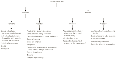
Causes of sudden vision loss in older persons include giant cell arteritis, stroke or transient ischemic attacks, ophthalmic artery or central retinal vein occlusion, acute angle-closure glaucoma, retinal detachment, and vitreous hemorrhage. Sudden vision loss most often stems from the abrupt evolution of one or more significant chronic medical problems.22,23
Some etiologies of sudden vision loss in older persons constitute ophthalmologic emergencies, and appropriate treatment necessitates a high index of suspicion and swift diagnosis. High-dose prednisone should be started immediately in suspected cases of giant cell arteritis; this can prevent vision loss, which is usually irreversible after the fact.24 Acute angle-closure glaucoma should be considered in any patient with severe headaches, nausea, vomiting, and visual acuity changes; confirmatory physical findings, such as acute red eye, cloudy cornea, and a sluggishly responsive, mid-dilated pupil, should be sought. Figure 2 will aid in the differential diagnosis of sudden vision loss in older persons.

TREATMENT OF EYE DISEASE IN PERSONS WITH DIABETES
The treatment of choice for persons with diabetes and severe retinopathy is panretinal laser photocoagulation. In severe proliferative diabetic retinopathy, panretinal laser photocoagulation reduces the risk of severe vision loss by at least 50 percent.25 Macular edema is treated with focal laser photocoagulation, which may prevent or delay vision loss in up to 70 percent of cases.25 Persons with diabetes are at increased risk of vitreous hemorrhage and retinal detachment. Specific treatment of these conditions is beyond the scope of this article.
TREATMENT OF AMD
Retinal photodynamic therapy may arrest progression in the neovascular form of AMD.2 Pegaptanib (Macugen) and ranibizumab (Lucentis) inhibit vascular endothelial growth factors involved in angiogenesis,26,27 and have shown promising results in treating neovascular AMD. Intravitreal injection is required, which may limit patient acceptance. The place of these agents and timing of their administration in AMD therapy is the subject of ongoing clinical trials.
TREATMENT OF PRIMARY OPEN-ANGLE GLAUCOMA
The most commonly administered ophthalmic preparations in older persons are for the treatment of primary open-angle glaucoma. The aim of these agents is to lower intraocular pressure in persons with ocular hypertension and evidence of optic nerve damage.28 Systemic absorption of these ophthalmic preparations can be reduced by nasolacrimal occlusion (finger pressed on the caruncle) and eyelid closure for 30 seconds after instillation of the drop.29 Topical ophthalmic agents used for the treatment of primary open-angle glaucoma are summarized in Table 5.30
| Drug class and agents | Mechanism of action | Selected drug interactions and effects | Precautions or warnings |
|---|---|---|---|
| Alpha-adrenergic agonists | |||
| Apraclonidine (Iopidine), brimonidine (Alphagan) | Decrease aqueous humor production | Monoamine oxidase inhibitors: contraindicated because of hypertensive urgency or emergency | Not studied in persons with hepatic or renal impairment |
| Beta-adrenergic antagonists | |||
| Betaxolol (Betoptic), carteolol, levobunolol (Betagan), metipranolol (Optipranolol), timolol (Betimol) | Decrease aqueous humor production | Calcium antagonists and digitalis: prolong atrioventricular conduction time | Asthma, cardiac conduction abnormalities, heart failure |
| Oral beta antagonists: hypotension and bradycardia | |||
| Carbonic anhydrase inhibitors | |||
| Brinzolamide (Azopt), dorzolamide (Trusopt Ocumeter) | Decrease aqueous humor production | No significant interactions reported | Sulfonamides: potential for Stevens-Johnson syndrome |
| Parasympathomimetics | |||
| Carbachol (Isopto Carbachol), pilocarpine | Increase aqueous humor outflow | Topical nonsteroidal anti-inflammatory drugs: unconfirmed reports of rendering carbachol ineffective | Gastric or urinary tract obstruction, peptic ulcer disease, asthma |
| Do not use in retinal disease | |||
| Prostaglandin analogues | |||
| Latanoprost (Xalatan), travoprost (Travatan), bimatoprost (Lumigan) | Increase aqueous humor outflow | Thimerosal: precipitation of latanoprost | Intraocular inflammation, macular edema |
| Sympathomimetics | |||
| Dipivefrin (Propine), epinephrine | Decrease aqueous humor production and increase aqueous humor outflow | Levothyroxine, antihistamines, tricyclic antidepressants: potential for systemic interaction with increased pressor response | Hypertension, coronary artery disease, known arrhythmia, diabetes, asthma (epinephrine) |
| Systemic effects rare with dipivefrin | |||
Prevention of Vision Loss
Aggressive management of chronic medical disorders in older persons can help preserve vision. Smoking cessation, limiting exposure to ultraviolet light, and (possibly) dietary changes and selected use of antioxidant or trace mineral supplements may preserve vision in older persons.
AGGRESSIVE BLOOD GLUCOSE CONTROL
Intensive control of blood glucose has been shown to reduce the progression of diabetic retinopathy in persons with type 1 and type 2 diabetes.25 A 10-year poststudy analysis of survivors in the U.K. Prospective Diabetes Study (UKPDS) showed a persistent and significant 24 percent decrease in relative risk of microvascular events in the intensive blood glucose control group, even if tight glucose control was subsequently lost.31 It is challenging to apply this evidence to older persons with diabetes, however, because older adults are particularly susceptible to hypoglycemia with tight glucose control.32 Therapy must be individualized to the patient; as a general rule, blood glucose in older persons with diabetes should be as tightly controlled as possible while avoiding hypoglycemia and its attendant risks.
BLOOD PRESSURE MANAGEMENT
Aggressive blood pressure control with a target of less than 150/85 mm Hg is likely to be vision preserving in older persons, especially those with diabetes. The UKPDS demonstrated that lowering blood pressure to below 150/85 mm Hg in persons with diabetes reduces the risk of progressive diabetic retinopathy, irrespective of A1C level.33 A 10 mm Hg decrease in systolic blood pressure provided an 11 percent relative risk reduction in the incidence of photocoagulation or vitreous hemorrhage; however, unlike intensive blood glucose control, blood pressure lowering must be sustained over time to preserve any benefit.34,35
Hypertension (with or without a diagnosis of diabetes) is associated with a higher risk of ischemic eye events, such as central retinal vein occlusion.36
LIPID MANAGEMENT
Hyperlipidemia is an independent risk factor for central retinal artery and vein occlusion.36,37 Observational studies suggest that control of hyperlipidemia in older persons with and without diabetes may independently reduce the risk of vision loss; however, this has not been confirmed in randomized controlled trials.1,3
SMOKING CESSATION
Smoking has been linked to a variety of causes of visual impairment in older persons, including AMD,38 cataracts,39 and progressive diabetic retinopathy.40 For ophthalmic health, as well as numerous other benefits, older persons who smoke should be advised to quit and offered smoking cessation counseling.
ULTRAVIOLET LIGHT EXPOSURE
Cumulative ultraviolet light exposure is linked to the development of cataracts.41 Older adults should be advised to consider the routine use of sunglasses that filter out ultraviolet light when driving or engaged in outdoor activity.
DIET AND SUPPLEMENTS
A Cochrane meta-analysis reviewed three large prospective clinical trials of antioxidant supplements or zinc to prevent or delay the onset of AMD, and found no demonstrable benefit.42
The Age-Related Eye Disease Study (AREDS) enrolled 3,640 persons with established AMD in a four-arm, randomized, prospective clinical trial of antioxidant supplementation, antioxidants plus zinc, zinc plus copper, and placebo. High-risk persons (those with more advanced disease at enrollment) randomized to the antioxidants plus zinc group had statistically significant preservation of vision compared with the placebo group (estimated odds reduction of 27 percent).43
Important caveats are attached to the use of these supplements. Excessive intake of vitamins A and E, especially in smokers, has been linked with an increased risk of lung cancer, and possibly higher rates of congestive heart failure.44,45 Reanalysis of the AREDS data suggests that zinc supplementation is associated with an increased risk of hospitalization for urologic problems.46
There are no prospective or randomized trials of anti-oxidant supplementation for prevention or treatment of eye diseases other than AMD. Observational studies have shown conflicting association between high levels of dietary intake of antioxidants and cataract formation.47
Routine antioxidant or mineral supplementation in all older adults for prevention of AMD or other eye diseases cannot yet be recommended until ongoing, prospective trials clarify who may benefit and what harms, if any, might result from long-term supplementation. There is encouraging evidence, which needs to be further validated, that specific antioxidant and zinc supplementation (the regimen used in AREDS) may preserve vision in some persons with advanced AMD.