Chronic wounds are those that do not progress through a normal, orderly, and timely sequence of repair. They are common and are often incorrectly treated. The morbidity and associated costs of chronic wounds highlight the need to implement wound prevention and treatment guidelines. Common lower extremity wounds include arterial, diabetic, pressure, and venous ulcers. Physical examination alone can often guide the diagnosis. All patients with a nonhealing lower extremity ulcer should have a vascular assessment, including documentation of wound location, size, depth, drainage, and tissue type; palpation of pedal pulses; and measurement of the ankle-brachial index. Atypical nonhealing wounds should be biopsied. The mainstay of treatment is the TIME principle: tissue debridement, infection control, moisture balance, and edges of the wound. After these general measures have been addressed, treatment is specific to the ulcer type. Patients with arterial ulcers should be immediately referred to a vascular surgeon for appropriate intervention. Treatment of venous ulcers involves compression and elevation of the lower extremities, plus exercise if tolerated. Diabetic foot ulcers are managed by offloading the foot and, if necessary, treating the underlying peripheral arterial disease. Pressure ulcers are managed by offloading the affected area.
A chronic wound is one that fails to progress through a normal, orderly, and timely sequence of repair, or in which the repair process fails to restore anatomic and functional integrity after three months.1 In 2014, wound care for Medicare beneficiaries cost an estimated $28 billion to $96.8 billion.2 A 2012 German study found a 1% to 2% prevalence of chronic nonhealing wounds in the general population.3 Chronic wounds, typically diabetic ulcers, preceded 85% of amputations.4 Some chronic wounds can take decades to heal, thus contributing to secondary conditions such as depression, and can ultimately lead to isolation and family distress. The five-year mortality rate after developing a diabetic ulcer is approximately 40%5; therefore, proper diagnosis and treatment of wounds and management of comorbidities are imperative.
SORT: KEY RECOMMENDATIONS FOR PRACTICE
| Clinical recommendation | Evidence rating | Comments |
|---|---|---|
| The ankle-brachial index may not be accurate in patients with diabetic foot ulcers; the toe-brachial index should be used instead to screen for peripheral arterial disease.14 | C | Disease-oriented study |
| Compression therapy has been proven beneficial for venous ulcer treatment and is the standard of care.29 | B | Cochrane review of lower-quality RCTs |
| Offloading with a total contact cast is the optimal treatment for diabetic foot ulcers.32,33 | A | Cochrane review of RCTs and a single RCT comparing treatment outcomes |
| A repositioning schedule should be established to treat and prevent pressure ulcers in hospitalized patients, and patients should not be positioned on bony prominences.36,37 | C | Consensus guidelines |
RCT = randomized controlled trial.
A = consistent, good-quality patient-oriented evidence; B = inconsistent or limited-quality patient-oriented evidence; C = consensus, disease-oriented evidence, usual practice, expert opinion, or case series. For information about the SORT evidence rating system, go to https://www.aafp.org/afpsort.
BEST PRACTICES IN WOUND CARE
| Recommendation | Sponsoring organization |
|---|---|
| Do not culture or treat clinically uninfected lower extremity wounds with systemic antibiotics. | American Podiatric Medical Association |
| Do not use whirlpools for wound management. | American Physical Therapy Association |
Source: For more information on the Choosing Wisely Campaign, see https://www.choosingwisely.org. For supporting citations and to search Choosing Wisely recommendations relevant to primary care, see https://www.aafp.org/afp/recommendations/search.htm.
Stages of Wound Healing
Wound healing is a complex sequence of events that begins with injury and ends with successful closure. It typically moves through four stages: hemostasis/coagulation, inflammation, proliferation, and maturation/remodeling (Table 1).6 These phases overlap as cytokines and growth factors guide the healing process. Chronic wounds most often do not progress past the inflammatory phase.
TABLE 1. Stages of Wound Healing
| Day | Stage | Pathophysiology | Outcome |
|---|---|---|---|
| 0 to 3 | Hemostasis/coagulation | Clotting, vasoconstriction | Bleeding stops |
| 1 to 25 | Inflammation | Cytokines and growth factors released; vasodilation; phagocytosis (i.e., white blood cells engulf bacteria and debris); neutrophils release reactive oxygen species and proteases | Redness, swelling, warmth, and pain |
| 1 to 25 | Proliferation | Collagen (type III) synthesis, granulation formation, epithelialization, angiogenesis, contraction | New blood vessel growth, wound closure |
| 20 to 365 | Maturation/remodeling | Scar remodeling (type III to I collagen) | Scar strength increases to approximately 80% of original tissue |
Information from reference 6.
Causes of Chronic Wounds
All wounds have the potential to become chronic wounds. They are classified by etiology into four categories, each with its own typical location, depth, and appearance: arterial, diabetic, pressure, and venous ulcers7 (Table 2). To appropriately treat a chronic wound, an understanding of the pathophysiology of the wound is critical. Despite differences in etiology, chronic wounds share certain features, including excessive levels of proinflammatory cytokines, persistent infections, formation of drug-resistant microbial biofilms, and senescent cells that do not respond to reparative stimuli.8
TABLE 2. Assessment of Chronic Wounds
| Wound type | Appearance | Assessment |
|---|---|---|
| Arterial ulcer | Deep; eschar; punched-out, well-demarcated borders; deep structures may be exposed | Palpate pulses |
| Assess macrocirculation: | ||
| Angiography | ||
| Ankle-brachial index or toe-brachial index | ||
| Lower extremity arterial duplex ultrasonography | ||
| Pulse volume recording | ||
| Segmental limb pressure | ||
| Assess microcirculation: | ||
| Skin perfusion pressure | ||
| Transcutaneous oximetry | ||
| Diabetic ulcer | Located on plantar aspect of foot, extensive callus formation, superficial to deep | Ankle-brachial index or toe-brachial index |
| Bone scan if concern for osteomyelitis | ||
| Lower extremity arterial duplex ultrasonography | ||
| Magnetic resonance imaging if concern for osteomyelitis | ||
| Radiography | ||
| Transcutaneous oximetry | ||
| Wound culture | ||
| Pressure ulcer | Located over bony prominences, superficial to deep | Computed tomography if indicated |
| Magnetic resonance imaging if concern for osteomyelitis | ||
| Nutritional support | ||
| Radiography | ||
| Wound culture | ||
| Venous ulcer | Shallow, no eschar; located over medial aspect of lower extremity (gaiter region) | Palpate pulses |
| Ankle-brachial index | ||
| Doppler ultrasonography | ||
| Lower extremity arterial duplex ultrasonography | ||
| Lower extremity venous duplex ultrasonography with reflux | ||
| Transcutaneous oximetry |
Assessment and Classification
Assessment of wounds should begin with a thorough physical examination. A more focused examination of the wound itself can then help guide treatment. The wound location, size, and depth; presence of drainage; and tissue type should be documented. Based on the location and appearance, most chronic wounds can be categorized by etiology, which allows for adequate workup and treatment recommendations.
Venous ulcers are the most common type of chronic wound. They are typically shallow and located on the medial supramalleolar aspect of the lower extremity (Figure 1). There may be classic signs of venous hypertension, including edema, hemosiderin staining, and lipodermatosclerosis. Workup should include assessment of arterial status with the ankle-brachial index (ABI) and palpation of pulses to rule out mixed arterial and venous disease and to ensure adequate perfusion before compression.9 If necessary, further workup with venous duplex ultrasonography with reflux and/or arterial duplex ultrasonography will help with the diagnosis.
FIGURE 1.
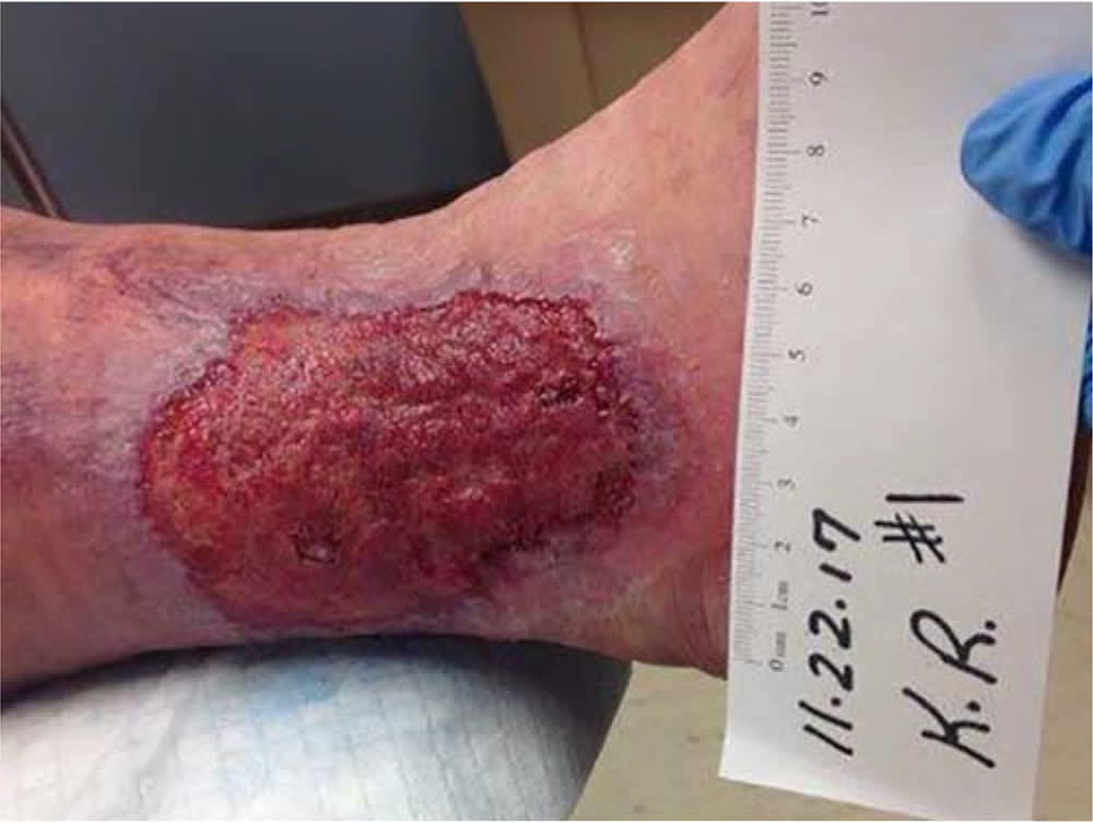
Venous ulcer.
Arterial ulcers are typically located on the distal extremities and may be deep, with tendon or bone exposed (Figure 2). As with venous ulcers, the initial workup should include ABI measurement and palpation of pulses. An ABI less than 0.8 may be a sign of arterial disease, and an ABI greater than 1.2 is consistent with noncompressible vessels. Both require further vascular assessment, which may include arterial Doppler/duplex ultrasonography, segmental limb pressures, pulse volume recording, skin perfusion pressure, or transcutaneous oximetry.10 Referral to a specialist should be considered if vascular test results suggest poor perfusion.
FIGURE 2.
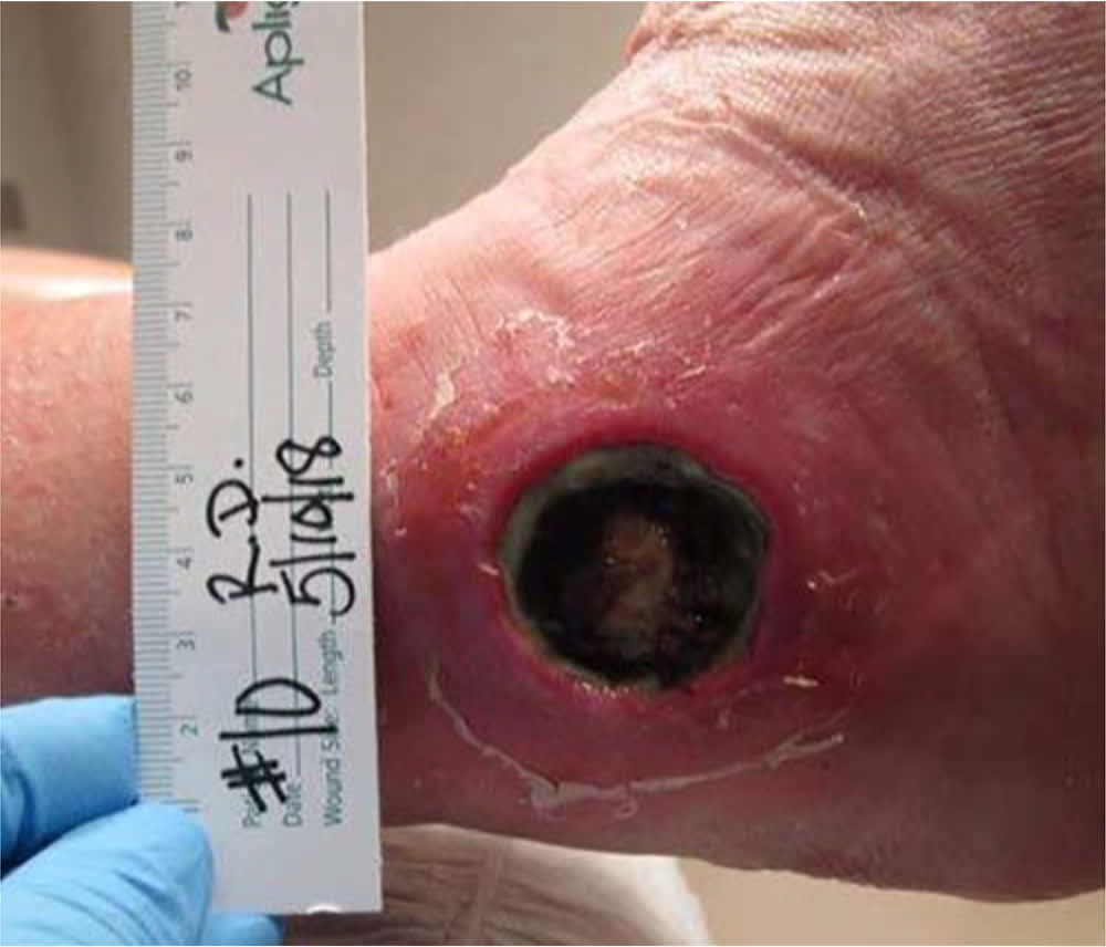
Arterial ulcer.
Diabetic ulcers are the most common cause of lower extremity amputation. Early intervention and management are essential given the high mortality rate after amputation.5 Diabetic foot ulcers are caused by a combination of underlying neuropathy, peripheral arterial disease, and structural deformities that cause increased pressure on affected areas of the foot.11 Diabetes mellitus affects sensory, motor, and autonomic nerve function. The combined effects result in structural deformities of the foot; dry, poorly hydrated integument; and an inability to detect pain and repetitive injury. Diabetes also causes higher rates of atherosclerotic disease.12 Diabetic ulcers are typically located on the toes or the plantar aspect of the metatarsal heads (Figure 3). They may have the characteristic crater-like appearance, be covered in eschar or necrotic tissue at the wound bed, and have exposed deep structures including tendon and bone. These ulcers can be shallow or deep and are usually surrounded by a thick ring of callus. Workup should include palpation of pedal pulses, ABI measurement,12 and assessment for neuropathy with Semmes-Weinstein monofilament.13 Imaging is often required to rule out deeper infection such as osteomyelitis. Patients with diabetes often have calcified, noncompressible arteries that result in an abnormally high ABI. A more reliable test in these patients is the toe-brachial index because the toe arteries rarely calcify.14
FIGURE 3.
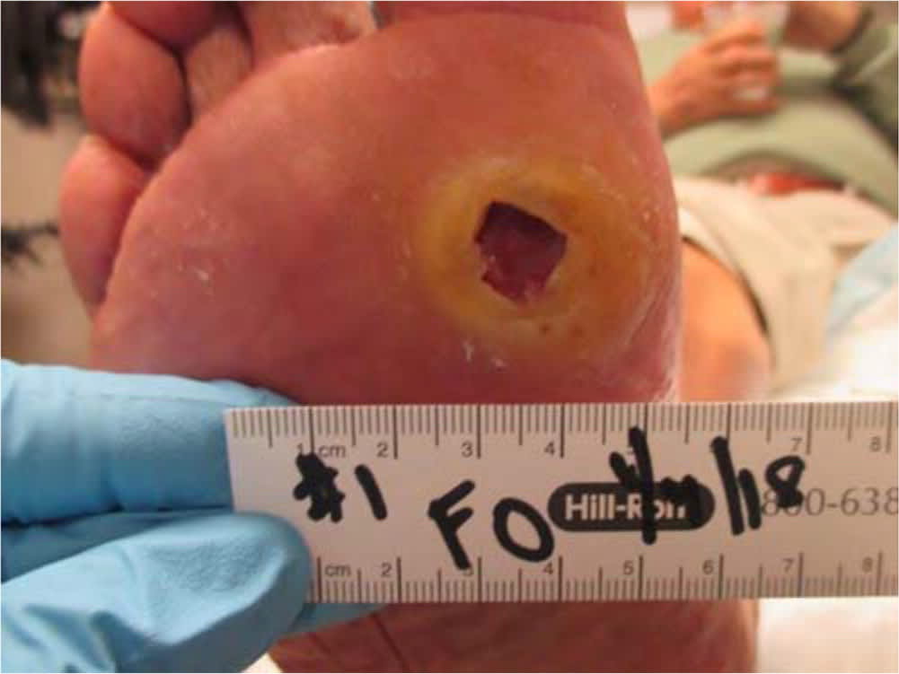
Diabetic foot ulcer.
The term pressure ulcer is preferred over bedsore, decubitus ulcer, or pressure sore. A pressure ulcer is a localized injury to the skin or underlying tissue—usually over a bony prominence such as the sacrum, coccyx, hip, or heel—that results from pressure in combination with shear force (Figure 4). Pressure ulcers can also be caused by devices such as nasal cannulas, nasogastric tubes, and casts or splints. Risk-assessment tools such as the Braden scale 15 can help minimize pressure ulcers by identifying high-risk patients. These ulcers are managed according to guidelines from the National Pressure Ulcer Advisory Panel, which classifies them into six stages based on the extent of tissue damage. Assessment may include a workup for infection, imaging to rule out deeper infection, and nutritional assessment.
FIGURE 4.
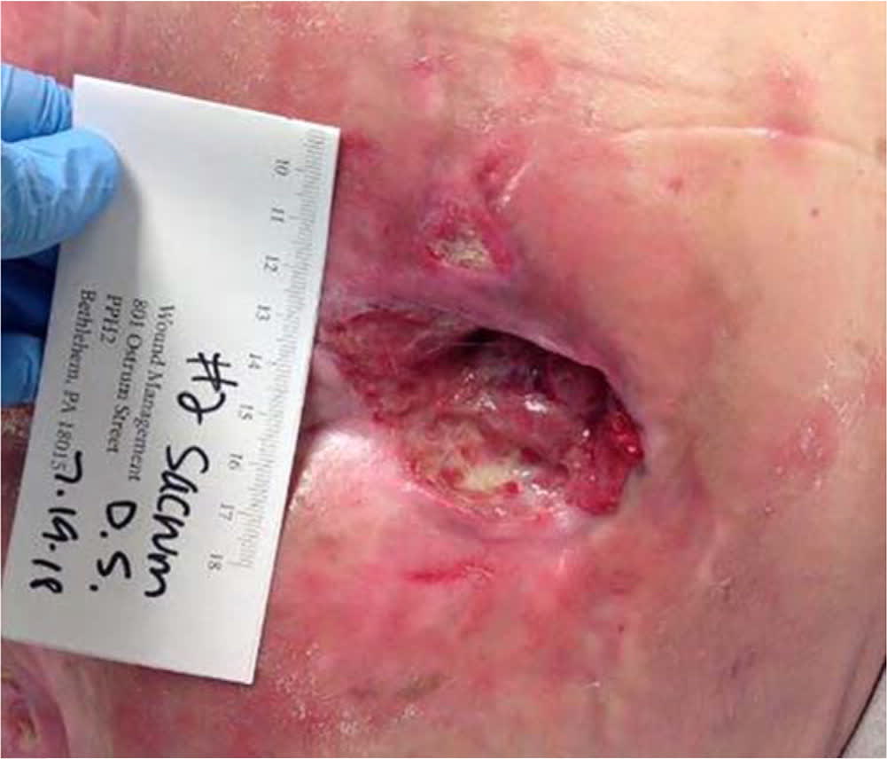
Stage 3 sacral pressure ulcer.
When assessing a chronic wound for infection, physicians should be aware that chronic infected wounds have different signs and symptoms than acute infected wounds. Typical signs of infection such as erythema, edema, pain, and fever are not always present. Two mnemonics are used to identify chronic wounds with infectious processes:
- NERDS (for wounds with biofilm or critical colonization): nonhealing, exudative, red and bleeds easily, debris, and smell. Sensitivity is 73% and specificity is 80.5% when three criteria are present.16
- STONEES (for infection): size increasing, temperature increased, os (probes to or exposed bone), new areas of breakdown, exudative, erythema/edema, and smell. Sensitivity is 90% and specificity is 69.4% when three criteria are present.16
Biopsy with quantitative processing is the best method of wound culture; a result of more than 100,000 colony-forming units is considered positive. Other options include the Levine and Z techniques. The Levine technique is superior, with several studies showing higher sensitivity and specificity compared with the Z technique.17 The Levine technique consists of debriding superficial necrotic tissue, then applying pressure with a swab while rolling it over 1 cm2 for five seconds to obtain fluid from deeper in the wound. In the Z technique, the handle of a swab is rotated between the fingers in a zig-zag fashion across the wound without touching the wound edge.
A chronic wound that does not respond to appropriate care may be an atypical wound. Atypical wounds are located in abnormal locations, have an abnormal appearance, and do not heal after three to six months of standard wound care. Atypical wounds have inflammatory, infectious, vasculopathic, metabolic, genetic, malignant, or external etiologies (e.g., calciphylaxis, pyoderma gangrenosum, vasculitis, autoimmune disease). Physicians should perform a biopsy for further evaluation in patients with suspected atypical wounds.18 Referral to a wound care specialist, dermatologist, or rheumatologist may also be indicated.
Treatment
Initially, all chronic wounds should be treated according to the TIME principle: tissue debridement (in all cases except arterial ulcers), infection control, moisture balance, and edges of the wound.19 After these general measures are addressed, the ulcer must be correctly diagnosed and classified so that appropriate care can be provided.
Debridement is the removal of dead cells and is an essential part of wound care. It is the first-line treatment for chronic wounds. A 2005 study showed that healing of chronic wounds is twice as likely with aggressive debridement.20 Another study of 154,664 patients with 312,744 wounds showed that consistent debridement at intervals of one week or less resulted in significantly faster healing and a 70.8% overall healing rate.21
In addition to removing dead cells and tissue, debridement of biofilm—multicellular communities held together by a self-produced extracellular matrix that can halt the healing process22—is a key factor in wound healing. The National Institutes of Health reports that 80% of wound infections are due to biofilm.23 Biofilm is invisible to the naked eye, and detection with different techniques is currently being investigated.24 Without destruction of biofilm, wound healing can stall during the inflammatory phase.25
There are several different types of debridement: surgical, autolytic, enzymatic, and biologic. Surgical debridement is used most often, but other methods may be appropriate depending on the type of wound. Infection is usually controlled with topical agents, including dressings with silver, polyhexamethylene biguanide, and cadexomer iodine. Antimicrobial washes may also be beneficial if biofilm is suspected. If cellulitis or signs of systemic infection are present, oral antibiotics may also be indicated.
Moisture balance is an essential part of wound care. Chronic wounds should never be exposed to air to “dry out,” as is often recommended. Moist wounds heal more quickly and have less risk of infection.26 If a wound appears dry, moisture needs to be added; this is accomplished by choosing an appropriate dressing (Table 3).6 Conversely, if a wound is draining, the drainage needs to be controlled and kept off of the periwound. The proper dressing should hold the moisture on the wound bed to prevent desiccation.
TABLE 3. Wound Dressings
| Wound appearance | Type of dressing |
|---|---|
| Draining | Calcium alginate |
| Collagen | |
| Composite | |
| Foam | |
| Hydrocolloid (for mild to moderate drainage) | |
| Infected/inflamed | Cadexomer iodine |
| Polyhexamethylene biguanide | |
| Silver impregnated | |
| Necrotic | Collagenase (enzymatic debridement) |
| Honey impregnated (autolytic debridement) | |
| Hydrocolloid (autolytic debridement) | |
| Maggots (biologic debridement) | |
| Nondraining/dry | Collagen gel |
| Composite | |
| Hydrocolloid | |
| Hydrogel | |
| Transparent film |
Information from reference 6.
Wound edges must be free from undermining. If edges are rolled, they should be excised. This allows for epithelialization, which is the clearest sign the wound is healing.27
TREATMENT BY WOUND ETIOLOGY
Venous Ulcers. Venous hypertension due to incompetent veins or venous obstruction distends the superficial veins, causing wall damage, exudation of fluid into the interstitial space, and edema of venous insufficiency. Treatment consists of exercise, which lowers venous pressures28; elevation of lower extremities; and adequate compression—usually 30 to 40 mm Hg—to improve venous return29 after vascular status has been proven adequate (Table 4).29,30 Patients may benefit from statin therapy for its vasoactive and anti-inflammatory effects.30 Early endovenous ablation may also be beneficial.31
TABLE 4. Treatment of Venous Ulcers
| Treatment | Comments |
|---|---|
| Compression29 | After vascular evaluation, compress 30 to 40 mm Hg as tolerated (contraindicated if ankle-brachial index < 0.6) |
| Stockings and wraps: | |
| Long stretch (> 140% extensibility) | |
| Short stretch (≤ 60% extensibility) | |
| Zinc oxide compression bandage (Unna Boot) | |
| Elevation | Elevation above level of the heart at rest |
| Exercise | Walking activates calf muscle pump |
| Simvastatin (Zocor)30 | Vasoactive and anti-inflammatory properties in addition to lipid-lowering properties |
| Clinically significant improvement in healing time and healing rates | |
| Wound care | TIME principle: tissue debridement, infection control, moisture balance, and edges of the wound |
Note: See recent review of venous ulcer treatments at https://www.aafp.org/afp/2019/0901/p298.html.
Arterial Ulcers. The first step in the management of arterial ulcers is treating the underlying cause, which may include vascular bypass, stents, or dilation by a vascular surgeon (Table 5). The goals of wound care before adequate perfusion is obtained are to prevent infection and minimize debridement. After vascular intervention, the TIME principle should be used.
TABLE 5. Treatment of Arterial Ulcers
| Treatment | Comments |
|---|---|
| Exercise | Planned graduated walking program |
| Intervention | Angiography, angioplasty, or stents |
| Vascular bypass surgery | |
| Wound care | TIME principle: tissue debridement, infection control, moisture balance, and edges of the wound |
| Apply emollients to keep skin pliable | |
| Vascular evaluation | |
| Healable (adequate perfusion): | |
| TIME | |
| Maintain moisture balance | |
| Nonhealable (ischemia): | |
| Control infection | |
| Limit debridement | |
| Maintain moisture balance |
Note: Compression is contraindicated for arterial ulcers.
Diabetic Ulcers. Treatment of diabetic ulcers begins with optimal control of blood glucose levels. Most patients with diabetic foot ulcers also have underlying peripheral arterial disease, which requires evaluation. The thick callus that typically surrounds diabetic foot ulcers requires surgical debridement. The preferred treatment approach for diabetic foot ulcers is offloading the foot to remove pressure from the affected area32,33 (Table 632–34 ). This can be accomplished with certain shoes or other devices, but the recommended method is a total contact cast.32,33 Some diabetic foot ulcers will benefit from hyperbaric oxygen therapy.35
TABLE 6. Treatment of Diabetic Ulcers
| Treatment | Comments |
|---|---|
| Compression | Evaluate for peripheral arterial disease |
| Compress 30 to 40 mm Hg as tolerated if edema is present and perfusion | |
| is adequate | |
| Exercise | Planned graduated walking program |
| Hyperbaric oxygen therapy34 | Significantly improves healing rate and quality of life |
| Offloading32,33 | Nonremovable offloading: |
| Plaster/fiberglass total contact cast | |
| Roll-on total contact cast (similar to fiberglass) | |
| Total contact cast with removable boot | |
| Removable offloading: | |
| Aircast pneumatic walker | |
| CAM walker | |
| Charcot restraint orthotic walker | |
| Half-wedge shoe | |
| Heel relief shoe | |
| Wound care | TIME principle: tissue debridement, infection control, moisture balance, and edges of the wound |
CAM = controlled ankle movement.
Pressure Ulcers. Localized treatment for pressure ulcers follows the same TIME principle used for all chronic wounds, but has a greater emphasis on pressure redistribution, offloading, and microclimate control6,36 (Table 76 ). A common mistake is the recommendation of donut-style cushions for offloading. These should not be used because they impede blood flow to the area and can actually worsen existing ulcers or increase the risk of developing a new ulcer.37
TABLE 7. Management of Pressure Ulcers
| Management | Comments |
|---|---|
| Appropriate movement of patient | Do not drag patient |
| Microclimate | Control moisture |
| Keep skin clean and dry | |
| Use barrier creams | |
| Wash with pH-balanced skin cleanser and water | |
| Nutritional support6 | Ensure adequate hydration |
| Increase protein intake to 1.5 g per kg body weight | |
| Supplements: | |
| Vitamin A | |
| Vitamin C | |
| Zinc | |
| Offloading | Change positions every one to two hours to limit pressure on affected area |
| Small shifts in weight every 15 minutes | |
| Pressure redistribution | Air- or fluid-filled mattress |
| Air- or gel-filled seat cushions | |
| No donut-style cushions | |
| Risk assessment | Braden scale |
| Wound care | TIME principle: tissue debridement, infection control, moisture balance, and edges of the wound based on wound stage |
Information from reference 6.
Data Sources: An initial PubMed search was performed using the key terms chronic wounds, diabetic foot ulcers, venous ulcers, arterial ulcers, and pressure ulcers. The search included clinical trials, systematic reviews, randomized controlled trials, and meta-analyses published since 1990. Several other searches were done using the Cochrane database, the Institute for Clinical Systems Improvement, the National Guideline Clearinghouse, and the Agency for Healthcare Research and Quality clinical guidelines and evidence reports. The Essential Evidence summary provided by AFP was also used, as were several landmark articles in wound care and guidelines from the major societies involved with wound care. Search date: May 2018.
The authors thank Jill Stoelzl, RN, CWOCN, and Elizabeth Shimer Bowers for editing and writing assistance.
