A 25-year-old woman presented with ulcerations of the lower extremities that developed one week earlier in the setting of hyperpigmentation over two to three years. She initially noticed multiple angular-shaped, erythematous nodules on the distal lower extremities that became hyperpigmented patches and plaques. The patient reported pain and itching in the involved areas. She had no history of diabetes mellitus or other chronic illnesses.
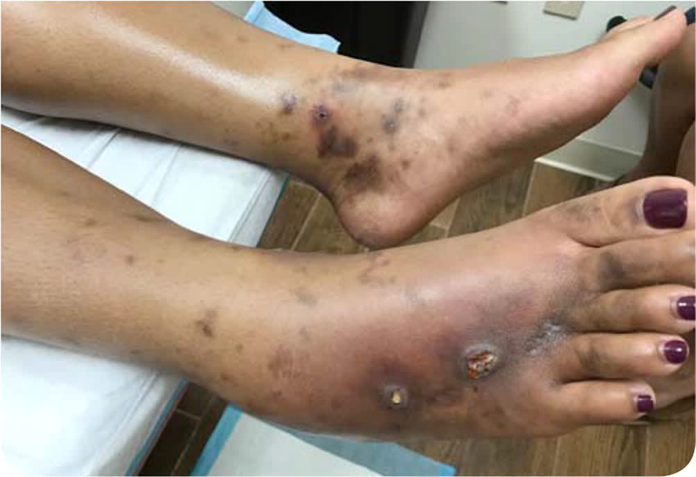
Physical examination revealed multiple tender, superficial ulcerations with surrounding erythema and hyperpigmentation on the medial aspect of the bilateral ankles and dorsum of her right foot (Figure 1). She also had an atrophic, white, stellate scar proximal to her right third and fourth metatarsophalangeal joints. Numerous purpuric patches and nodules were noted throughout the distal lower extremities. Her physical examination was otherwise unremarkable, and her vital signs were normal.
FIGURE 1.
Question
Based on the patient's history and physical examination findings, which one of the following is the most likely diagnosis?
A. Chronic venous insufficiency.
B. Cutaneous leukocytoclastic vasculitis.
C. Erythema nodosum.
D. Livedoid vasculopathy.
E. Pyoderma gangrenosum.
Discussion
The correct answer is D: livedoid vasculopathy. Livedoid vasculopathy is a chronic thromboocclusive cutaneous disorder marked by recurrent skin findings affecting the lower extremities. Characteristic clinical features are the initial appearance of tender or pruritic erythematous nodules, followed by superficial ulcerations and atrophic ivory-white scars with surrounding hyperpigmentation known as atrophie blanche.1 Ankle edema and peripheral purpura or petechiae may precede skin manifestations. Physical findings are often bilateral and most commonly observed on the distal lower extremity, ankle, or dorsal foot.2 Ulcerations are often painful and may persist for months despite treatment.
Livedoid vasculopathy most commonly presents in women with bimodal peaks at 45 to 50 and 70 to 75 years of age.1 Clinical findings are caused by the occlusion of cutaneous blood vessels by thrombi and fibrin due to hypercoagulability and impaired fibrinolysis.3 The clinical diagnosis can be confirmed with a 4- to 6-mm punch biopsy. Histopathology demonstrates intraluminal thrombi, endothelial proliferation, and connective tissue degeneration.3 Additional laboratory evaluation should be performed to evaluate for systemic rheumatic disorders. MTHFR mutations have been associated with this disease.
Treatment is focused on wound care and limiting further thrombi deposition through the use of antiplatelet medications and anticoagulants. Aspirin, 325 mg once daily, is the preferred initial treatment. For refractory cases, anticoagulation with warfarin (Coumadin) or low-molecular-weight heparin can be used for thromboembolism prophylaxis.4 The chronic nature of livedoid vasculopathy may cause the patient significant psychological and physical impairment.5
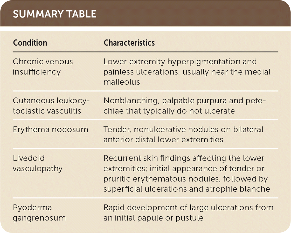
Chronic venous insufficiency often presents with lower extremity hyperpigmentation and painless ulcerations, usually near the medial malleolus. It is most common at older ages, with a peak prevalence in women 60 to 80 years of age.6 Venous ultrasonography may be useful in distinguishing venous insufficiency from livedoid vasculopathy.
Cutaneous leukocytoclastic vasculitis presents with non-blanching, palpable purpura and petechiae, indicating small vessel vasculitis. Cutaneous leukocytoclastic vasculitis does not typically cause ulcerations.
Erythema nodosum presents with tender, nonulcerative nodules on bilateral anterior distal lower extremities. It typically develops over several days and is often preceded by fever and other constitutional symptoms. It is most common in women between 25 and 54 years of age.7
Pyoderma gangrenosum presents with rapid development of large ulcerations from an initial papule or pustule. Patients have severe pain out of proportion to physical examination findings.
SUMMARY TABLE
| Condition | Characteristics |
|---|---|
| Chronic venous insufficiency | Lower extremity hyperpigmentation and painless ulcerations, usually near the medial malleolus |
| Cutaneous leukocytoclastic vasculitis | Nonblanching, palpable purpura and petechiae that typically do not ulcerate |
| Erythema nodosum | Tender, nonulcerative nodules on bilateral anterior distal lower extremities |
| Livedoid vasculopathy | Recurrent skin findings affecting the lower extremities; initial appearance of tender or pruritic erythematous nodules, followed by superficial ulcerations and atrophie blanche |
| Pyoderma gangrenosum | Rapid development of large ulcerations from an initial papule or pustule |
The opinions and assertions contained herein are the private views of the authors and are not to be construed as the official policy or position of the U.S. military, the Department of Defense, or the U.S. government.
