
Am Fam Physician. 2023;108(1):89-90
Author disclosure: No relevant financial relationships.
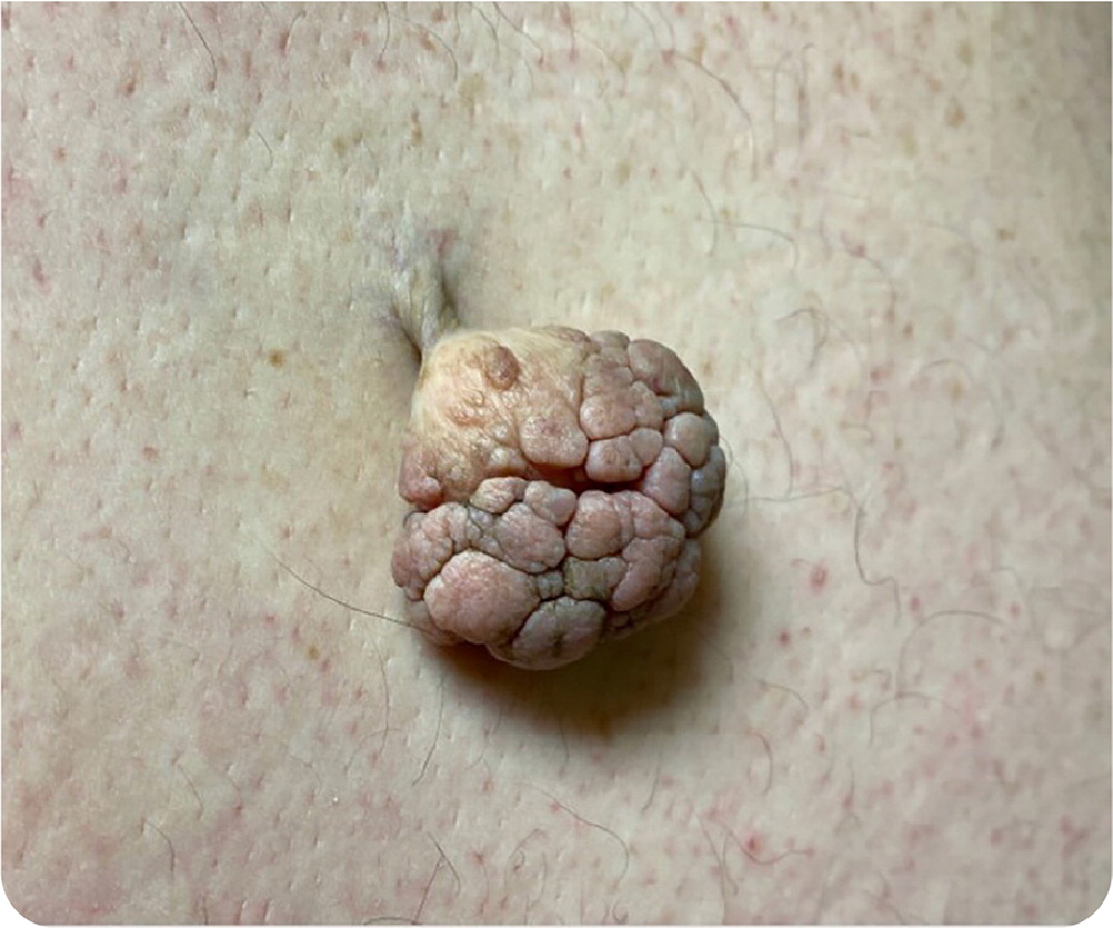
A 45-year-old man presented with a large skin growth on his back. The growth had been gradually enlarging since he first noticed it four years earlier. He had multiple similar, but smaller, growths around his eyes, neck, and upper back. His medical history included morbid obesity, hypertension, and recurrent gout.
Physical examination confirmed a pedunculated growth below his right scapula. The cauliflower dome was 3 cm in diameter, supported on a 1-cm stalk (Figure 1).
Question
Based on the patient's history and physical examination findings, which one of the following is the most likely diagnosis?
A. Dermatofibroma.
B. Dermatosis papulosa nigra.
C. Fibroepithelial polyp.
D. Seborrheic keratosis.
Discussion
The answer is C: fibroepithelial polyp. Also known as acrochordon or common skin tag, fibroepithelial polyp is a benign skin lesion of mesenchymal and ectodermal origin. Fibroepithelial polyps are often seen during routine physical examination as small, slightly discolored, papillomatous lesions, often in skin creases and areas exposed to friction. Rarely, large (greater than 5 mm), pedunculated polyps can be seen on the trunk.1
The prevalence increases with age; 50% to 60% of fibroepithelial polyps occur in adults older than 50 years. The formation of fibroepithelial polyps is not fully understood. Frequent skin rubbing and changes in epithelial sensitivity to hormones are proposed etiologies. Hormonal changes in conditions such as pregnancy, obesity, and diabetes mellitus may be associated with polyp growth.1–3
Smaller lesions (less than 5 mm) can be treated with cryotherapy or cauterization. Surgical excision is required for larger polyps.2
Dermatofibroma is a benign proliferation of fibroblasts appearing as a firm, raised, tan to reddish-brown papule or nodule, usually on the legs. Lesions are typically 3 to 10 mm in diameter and produce the dimple sign when laterally squeezed. The patient may report a history of local trauma (e.g., insect bite) or subcutaneous puncture (e.g., splinter).4,5
Dermatosis papulosa nigra is a benign skin condition characterized by pigmented filiform papules in the malar or temple region, neck, and upper trunk of individuals with darker skin tones. Lesions can be 1 to 5 mm in diameter, with a 1- to 3-mm elevation. Dermatosis papulosa nigra has a male to female ratio of 1: 2.6
Seborrheic keratosis is a benign skin growth of immature keratinocytes. It is the most common benign skin tumor in the older population. The yellow to brown lesions are highly pigmented, sharply demarcated, and soft to the touch. They may feel slightly greasy, velvety, or warty. They are commonly described as waxlike with a “stuck on” appearance.4
| Condition | Characteristics |
|---|---|
| Dermatofibroma | Firm, raised, tan to reddish-brown papule or nodule, usually on the legs; 3 to 10 mm in diameter; produces the dimple sign when laterally squeezed. |
| Dermatosis papulosa nigra | Pigmented filiform papules, commonly seen on malar or temple region, neck, and upper trunk of individuals with darker skin tones |
| Fibroepithelial polyp | Small, slightly demarcated, papillomatous lesion often seen in skin creases and areas exposed to friction |
| Seborrheic keratosis | Highly pigmented, sharply demarcated, soft to the touch; waxlike with a “stuck on” appearance |
