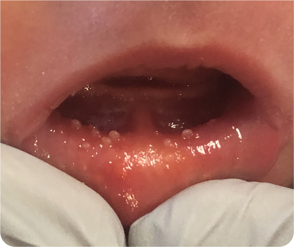
A 3,200-g infant was born at 37 weeks of gestation via repeat cesarean delivery to a woman (gravida 5, para 5) presenting with spontaneous rupture of membranes. The infant appeared healthy with Apgar scores of 8 and 9 at one and five minutes, respectively. No significant maternal history, prodromal symptoms, or abnormal examination findings were noted on admission. Regular prenatal care and testing were unremarkable. The parents denied a history of herpes simplex, but serology testing had not been performed. During establishment of breastfeeding in the first 24 hours of life, several small (1 to 3 mm) lesions were discovered on the mucosal surface of the newborn's lower lip.
Eversion of the infant's lower lip revealed nonfriable, white papules with minimal erythema at the base, intermixed with lesions of more vesicular appearance. No significant pain or discomfort was elicited on palpation. Mucoid, white material could be expressed when scraping pressure was applied to an isolated lesion. The newborn's physical examination was otherwise unremarkable, and his vital signs were within normal limits.
FIGURE 1
Question
Based on the patient's history and physical examination findings, which one of the following is the most likely diagnosis?
- A. Bohn nodules.
- B. Condyloma acuminata.
- C. Herpes simplex.
- D. Mucocele.
- E. Sebaceous hyperplasia.
Discussion
The answer is E: sebaceous hyperplasia. Intraoral, ectopic sebaceous gland hyperplasia (Fordyce spots) is a benign condition. It is characterized by isolated or clustered, white to yellow, fluid-filled vesicles, often with central umbilication. The lesions consist of sebaceous lobules, each containing clusters of sebocytes and a sebaceous duct in the submucosa or dermis.1 The lesions are not associated with hair follicles. Intraoral sebaceous hyperplasia occurs in approximately 1% of newborns and is believed to be caused by increased maternal androgen levels.1,2 In adults and older children, Fordyce spots are commonly seen in the mucosa of the lips and buccal area but can also occur on the gums and genital mucosa.3,4 Lesions are typically asymptomatic, nonfriable, and approximately 1 to 4 mm in size. Congenital sebaceous hyperplasia usually resolves spontaneously within a few weeks of birth; therefore, no treatment is indicated.1–3
Bohn nodules are asymptomatic, white to yellow, 2- to 3-mm, firm papules. They are found on the buccal and lingual alveolar ridge and are common in newborns. They have been called dental lamina cysts when located on the alveolar crests and Epstein pearls when located on the palate.5 Bohn nodules are keratin-filled cysts that spontaneously resolve within the first three months of life and require no treatment.6
Condyloma acuminata are attributed to in utero or post-natal human papillomavirus infection. The condition rarely presents at birth or early in the neonatal period due to a long latency period of six months to two years. Lesions are typically found in the anogenital region as multiple 1- to 5-mm, soft, filiform or flat-topped, verrucous papules that are the same color as surrounding skin. They can be discrete or clustered.5
Neonatal herpes simplex may present as erythematous macules, isolated or grouped vesicles, or a widespread vesiculobullous eruption that may include the buccal mucosa. Herpes simplex vesicles are characteristically clear, tender, and easily friable, with surrounding erythema. The vesicles may progress to a pustular, crusted, or ulcerated appearance. The lesions most commonly occur on the scalp and face or the presenting area in cases of breech positioning.7
Mucoceles are soft, round, translucent, 2- to 10-mm lesions on the buccal mucosa of the lower lip caused by trauma or obstruction of the minor salivary ducts. They are uncommon in newborns and are typically solitary.8 If hemorrhage into the mucocele has occurred, there may be a bluish hue. Lesions are painless, fluctuant, and tense.4
SUMMARY TABLE
| Condition | Examination findings | Other characteristics |
|---|---|---|
| Bohn nodules | Asymptomatic, white to yellow, 2- to 3-mm, firm papules; keratin-filled cysts; located on the buccal and lingual alveolar ridge | Common in newborns, with self-resolution by three months of life |
| Condyloma acuminata | Multiple 1- to 5-mm, soft, filiform or flat-topped, verrucous papules that are the same color as surrounding skin; lesions can be discrete or clustered | Attributed to in utero or postnatal human papillomavirus infection; rarely presents at birth or early in the neonatal period due to a long latency period of six months to two years |
| Herpes simplex | Tender vesicular lesions on an erythematous base; may present at various stages of healing | Erythematous macules, isolated or grouped vesicles, or widespread vesiculobullous eruption; may include the buccal mucosa; clear, tender, easily friable, with surrounding erythema |
| Mucocele | Painless, soft, round, translucent, 2- to 10-mm lesion on the buccal mucosa of the lower lip; usually solitary | Caused by trauma or obstruction of the minor salivary ducts; uncommon in newborns |
| Sebaceous hyperplasia | Asymptomatic, isolated or clustered, white to yellow, fluid-filled vesicles, often with central umbilication | Self-limited, resolving within a few weeks to months with no treatment |
The views expressed in this work are those of the authors and do not reflect the official policy or position of the U.S. military, the U.S. Department of Defense, or the U.S. government.
