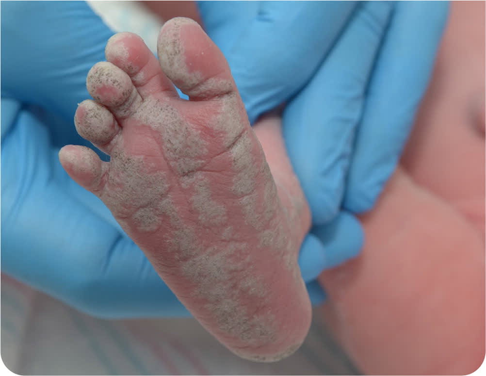
A female infant born at 39 1/7 weeks of gestation via uncomplicated spontaneous vaginal delivery was evaluated in the delivery room. Her Apgar scores were 9/9. Physical examination revealed dry, whitish-gray plaques in a linear pattern from the right inguinal area over the medial thigh and lateral shin, involving the plantar and dorsal surfaces of the right foot (Figure 1 and Figure 2). The remainder of the physical examination was unremarkable. The patient was afebrile with a normal pulse and respiratory rate. Laboratory test results were within normal limits, including routine newborn screenings and complete blood count.
FIGURE 1
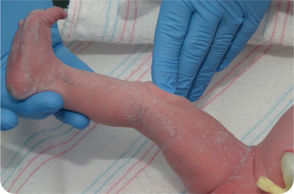
FIGURE 2
Question
Based on the patient's history and physical examination findings, which one of the following is the most likely diagnosis?
- A. Congenital herpes simplex.
- B. Congenital syphilis.
- C. Inflammatory linear verrucous epidermal nevus.
- D. Sebaceous nevus.
Discussion
The answer is C: inflammatory linear verrucous epidermal nevus (ILVEN), a variant of an epidermal nevus or mole. It is characterized by recurrent inflammation with a chronic eczematic or psoriasiform component. ILVEN is a benign overgrowth of cells in the epidermis that presents as erythematous, verrucous papules and plaques that characteristically follow the lines of normal embryologic cell migration in the skin (Blaschko lines). The lesions can be red, brown, or the same color as the surrounding skin.1 ILVEN is commonly found on the lower extremities. Children are most often affected, although cases of adult-onset ILVEN have been reported.2
ILVEN has a female predominance. Although most cases are sporadic, the condition may have a genetic component.3,4 The pathophysiology of the condition is unclear; however, it is believed to result from genetic mosaicism caused by somatic mutations.5 The classic diagnostic criteria for ILVEN are (1) unilateral, verrucous, intensely pruritic lesions following a linear distribution, (2) early age of onset, and (3) lesions that are refractory to treatment.6
Therapeutic options for ILVEN include topical steroids, fluocinonide ointment, tacrolimus, surgical excision, and carbon-dioxide laser therapy, although ILVEN is typically resistant to treatment.7,8 There is no definitive cure, but remission has been reported.9,10
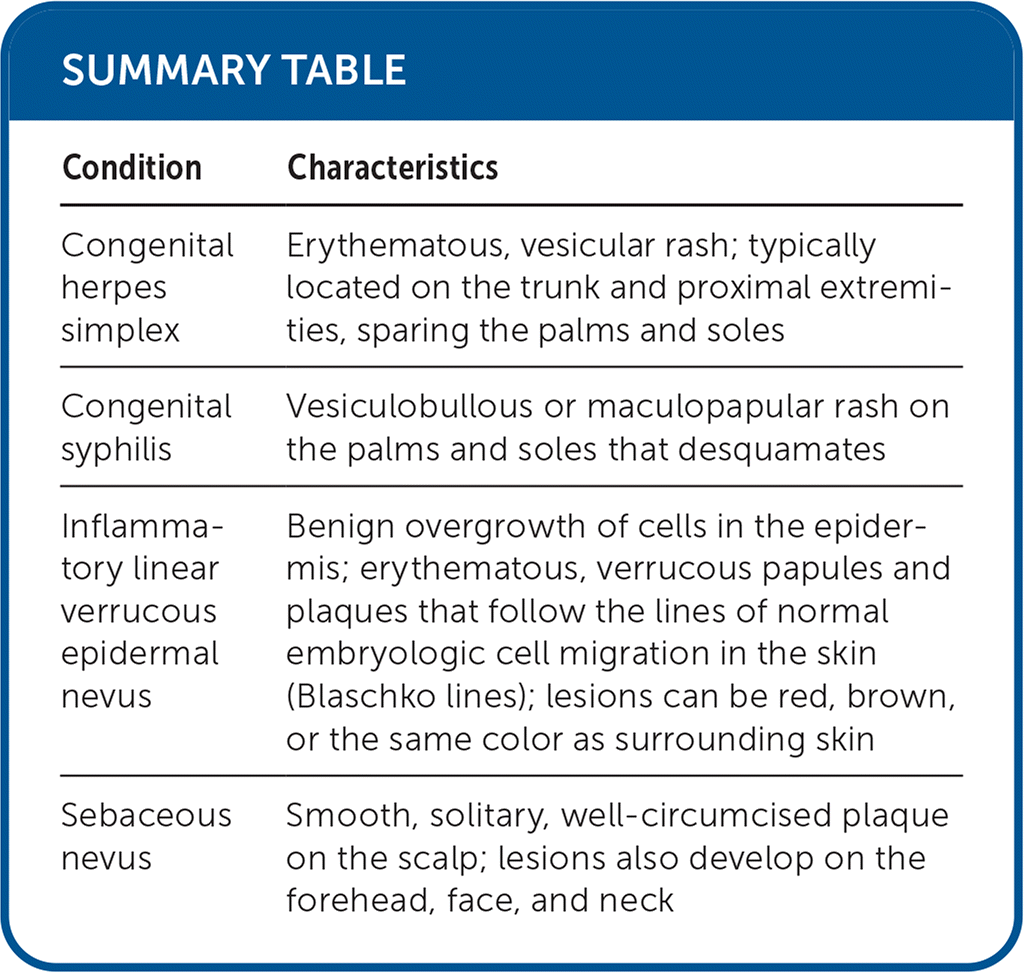
Congenital herpes simplex is most commonly transmitted with the passage of the newborn through an infected maternal genital tract, although ascending infection to the fetus in utero is possible.11 Newborns with congenital herpes simplex can present with irritability; lethargy; an erythematous, vesicular rash; and encephalitis. The rash is typically located on the trunk and proximal extremities, sparing the palms and soles.
Congenital syphilis is caused by Treponema pallidum, usually transmitted through ascending infection to the fetus in utero. Symptoms include copious mucopurulent nasal discharge, hepatosplenomegaly, and skeletal malformations. In newborns, dermatologic manifestations include a vesiculobullous or maculopapular rash on the palms and soles that desquamates.12
Sebaceous nevus is a congenital malformation of the pilosebaceous follicular unit. It typically presents at birth or shortly after and is characterized by a smooth, solitary, well-circumcised plaque on the scalp. Lesions also develop on the forehead, face, and neck. These lesions often become more verrucous and prominent in adolescence.13
SUMMARY TABLE
| Condition | Characteristics |
|---|---|
| Congenital herpes simplex | Erythematous, vesicular rash; typically located on the trunk and proximal extremities, sparing the palms and soles |
| Congenital syphilis | Vesiculobullous or maculopapular rash on the palms and soles that desquamates |
| Inflammatory linear verrucous epidermal nevus | Benign overgrowth of cells in the epidermis; erythematous, verrucous papules and plaques that follow the lines of normal embryologic cell migration in the skin (Blaschko lines); lesions can be red, brown, or the same color as surrounding skin |
| Sebaceous nevus | Smooth, solitary, well-circumcised plaque on the scalp; lesions also develop on the forehead, face, and neck |
