A 38-year-old woman presented with a painful mass in her right breast that had been present for a few months. She was six months postpartum, following an uncomplicated pregnancy and delivery. The patient did not breastfeed/chestfeed but reported continued lactation from her right breast. She had a history of fibroadenoma in the right breast, which was biopsied 15 years prior.
Physical examination revealed a large retroareolar mass that was mobile and fluctuant. The patient did not have skin changes or axillary or supraclavicular lymphadenopathy, and milky white fluid could be expressed from the right nipple. The left breast was normal, with no discharge. Laboratory workup showed an elevated prolactin level and a normal thyroid-stimulating hormone level.
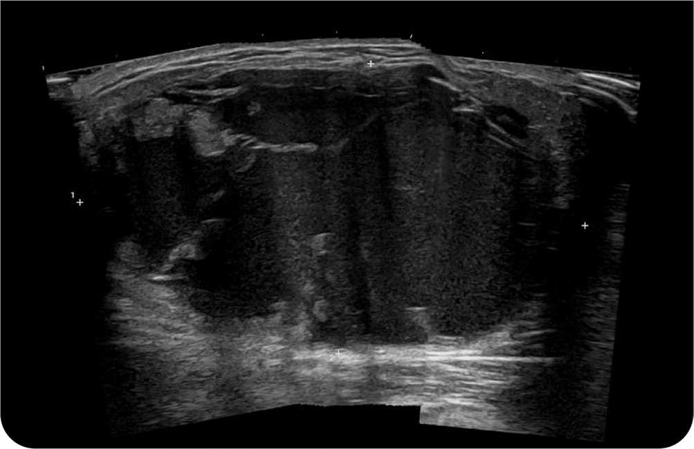
Diagnostic mammography and breast ultrasonography demonstrated a mixed attenuation, multiloculated cystic mass with flat fluid levels in the retroareolar aspect of the right breast (Figure 1 and Figure 2). The mass measured 6.9 × 6.2 × 6 cm. Magnetic resonance imaging of the head did not show intracranial abnormalities.
FIGURE 1
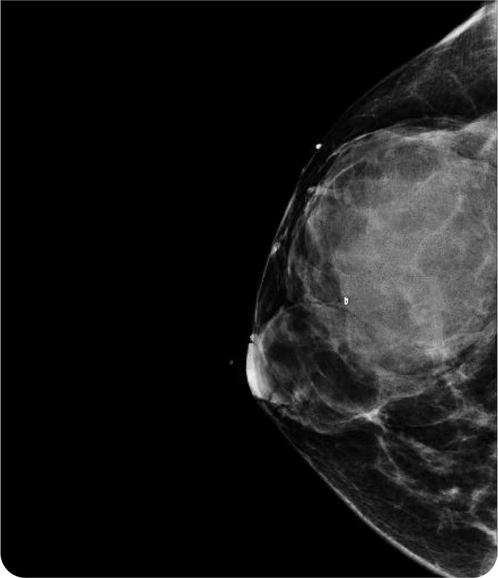
FIGURE 2
Question
Based on the patient's history, physical examination, and imaging findings, which one of the following is the most likely diagnosis?
- A. Abscess.
- B. Carcinoma.
- C. Cyst.
- D. Fibroadenoma.
- E. Galactocele.
Discussion
The answer is E: galactocele, a benign, milk-filled retention cyst that occurs during lactation. Galactoceles develop from duct obstruction.1 They can occur anywhere in the mammary system but are typically found in the retroareolar region of the breast.1 They are usually solitary and painless. A galactocele is typically small when first noticed but can become large over time.
The condition is diagnosed with breast ultrasonography and fine-needle aspiration of the white milky fluid.1 Aspiration is both diagnostic and therapeutic. This patient was referred to a breast surgeon who performed ultrasound-guided aspiration of 80 mL of milk.
Breast abscesses are localized, pus-filled, fluctuant masses in the breast tissue, with surrounding erythema and systemic symptoms, such as fever. They are usually a result of untreated or undertreated mastitis or cellulitis. Treatment comprises incision and drainage with antibiotic therapy.2,3
Breast carcinoma is the second most common cancer in women.3 In the absence of screening, breast carcinoma typically presents as a solid, immobile mass. It is diagnosed through imaging and biopsy. Treatment includes a combination of surgery, chemotherapy, and radiation therapy.
Breast cysts are benign, fluid-filled, fluctuant sacs in breast tissue. Many resolve spontaneously. For large or uncomfortable cysts, aspiration and drainage can relieve symptoms.3,4
Fibroadenoma is the most common noncancerous breast mass. It commonly occurs in patients between 15 and 35 years of age. The masses are often firm, painless, and unilateral and tend to get smaller to the point of resolution with age. Diagnosis is made by ultrasonography. Surgical excision can be performed if the mass causes symptoms or does not resolve.3,4
SUMMARY TABLE
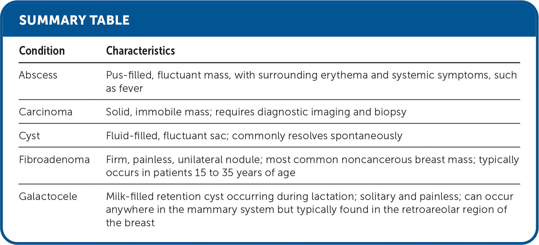
| Condition | Characteristics |
|---|---|
| Abscess | Pus-filled, fluctuant mass, with surrounding erythema and systemic symptoms, such as fever |
| Carcinoma | Solid, immobile mass; requires diagnostic imaging and biopsy |
| Cyst | Fluid-filled, fluctuant sac; commonly resolves spontaneously |
| Fibroadenoma | Firm, painless, unilateral nodule; most common noncancerous breast mass; typically occurs in patients 15 to 35 years of age |
| Galactocele | Milk-filled retention cyst occurring during lactation; solitary and painless; can occur anywhere in the mammary system but typically found in the retroareolar region of the breast |
