

Lymphadenopathy occurs in 0.6% of the population annually, usually from a benign cause. History should focus on lymph node location, duration of lymphadenopathy, associated symptoms (particularly fever, night sweats, and unintentional weight loss), past and current medical conditions, occupation, travel history, animal exposures, medication use, recent vaccine history, drug use, sexual history, and family history. Physical examination should first differentiate localized from generalized lymphadenopathy. Generalized lymphadenopathy is usually caused by underlying systemic disease. Although usually benign, localized lymphadenopathy may represent infection or malignancy, particularly if epitrochlear or supraclavicular nodes are affected. Lymph nodes that are larger than 2 cm, hard, or matted/fused to surrounding structures may indicate malignancy or granulomatous diseases, especially in children. When lymphadenopathy persists beyond four weeks or is accompanied by systemic symptoms, imaging and appropriate laboratory studies (eg, complete blood cell count, C-reactive protein, erythrocyte sedimentation rate, tuberculosis testing) should be obtained. Biopsy may be performed through fine-needle aspiration, core needle biopsy, or excisional biopsy. Antibiotics may be considered if a benign or self-limiting etiology (eg, bacterial lymphadenitis) is suspected. Corticosteroids should be avoided because they can mask the histologic diagnosis of lymphoma or other malignancy.
Lymphadenopathy is defined as lymph nodes with abnormal growth or character.1 The estimated annual incidence of lymphadenopathy is 6 in 1,000 people.2 The etiology can be infectious or noninfectious.2–6 Due to the extensive differential diagnosis for lymphadenopathy, which includes serious conditions such as lymphoma, autoimmune disorders, HIV, and metastatic cancer, taking a stepwise approach to evaluation is important.2–7
SORT: KEY RECOMMENDATIONS FOR PRACTICE
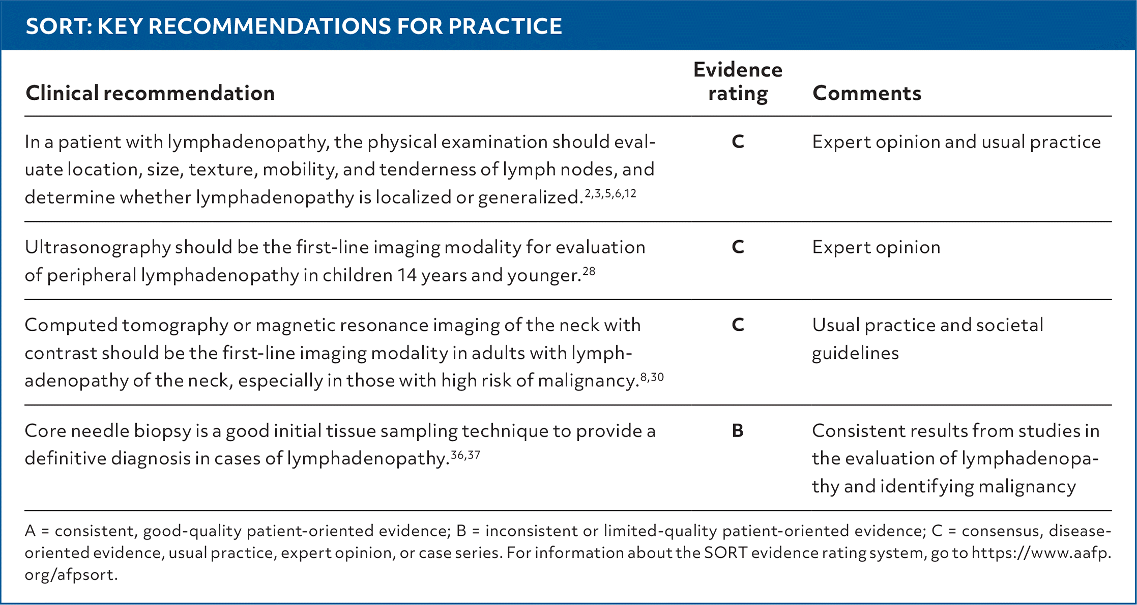
| Clinical recommendation | Evidence rating | Comments |
|---|---|---|
| In a patient with lymphadenopathy, the physical examination should evaluate location, size, texture, mobility, and tenderness of lymph nodes, and determine whether lymphadenopathy is localized or generalized.2,3,5,6,12 | C | Expert opinion and usual practice |
| Ultrasonography should be the first-line imaging modality for evaluation of peripheral lymphadenopathy in children 14 years and younger.28 | C | Expert opinion |
| Computed tomography or magnetic resonance imaging of the neck with contrast should be the first-line imaging modality in adults with lymphadenopathy of the neck, especially in those with high risk of malignancy.8,30 | C | Usual practice and societal guidelines |
| Core needle biopsy is a good initial tissue sampling technique to provide a definitive diagnosis in cases of lymphadenopathy.36,37 | B | Consistent results from studies in the evaluation of lymphadenopathy and identifying malignancy |
A = consistent, good-quality patient-oriented evidence; B = inconsistent or limited-quality patient-oriented evidence; C = consensus, disease-oriented evidence, usual practice, expert opinion, or case series. For information about the SORT evidence rating system, go to https://www.aafp.org/afpsort.
In the primary care setting, lymphadenopathy is caused by malignancy in 1.1% of cases overall, but in approximately 4% of individuals older than 40 years.2 Although lymphadenopathy usually has an easily identifiable cause, some patients present with an enlarged lymph node as the sole symptom or with nonspecific symptoms (eg, fatigue), making diagnosis challenging.3,7 In these situations, laboratory tests, imaging, or biopsy may be required.5,8 The MIAMI mnemonic (malignancies, infections, autoimmune disorders, miscellaneous/unusual conditions, and iatrogenic causes) is a helpful tool in assessing the differential diagnosis of lymphadenopathy (Table 1).6,9–11
Read the full article
Get immediate access, anytime, anywhere.
Choose a single article, issue, or full-access subscription.
Earn up to 14 CME credits per issue.
