

Active children and adolescents have unique risk factors for musculoskeletal injuries compared with adults. Physes and developing bones are at higher risk of injury than tendons and ligaments. Children’s bone remodeling is robust, allowing most clavicle fractures and torus fractures of the forearm to be managed conservatively. Radial head subluxation is managed with reduction. Apophyseal injuries are traction or overuse injuries that typically can be managed nonoperatively. Osteochondritis dissecans and other osteochondroses require frequent monitoring and occasionally surgical intervention.
Case 4. AJ is a 14-year-old patient who is brought to your office by her parents for pain and swelling in the right ankle 2 days after an inversion injury during a basketball game. After the injury, AJ stopped playing, was unable to bear weight, and hopped to the bench on her left foot with assistance from her teammates. She has been icing and elevating the ankle with some improvement, but she is afraid to walk on it.
Ankle Injuries
Ankle injuries in patients younger than 18 years are common. The incidence in males peaks between ages 15 and 19 years, whereas incidence in females peaks between ages 10 and 14 years.120 Males fracture their ankles 2 times as frequently as females.121 In children and adolescents, the presence of a physis contributes to unique fracture patterns that the family physician must be aware of to properly manage the injury.
ANATOMY
The true ankle joint consists of the tibia, fibula, and talus.122 Laterally, the joint is stabilized by the anterior talofibular ligament (ATFL), the posterior talofibular ligament (PTFL), and the calcaneofibular ligament (CFL) (Figure 7). Medially, the joint is stabilized by the multicomponent deltoid ligament. The tibiofibular syndesmosis provides further stability, running the length of the tibia and fibula, and distally comprises a complex of ligaments that reinforce the distal portion of the tibia to the fibula (Figure 8). These distal components attach to the epiphyses.121 Growing children with open physes can have isolated or combined avulsion, physeal, or ligamentous injury. Bony anatomy can be visualized with anteroposterior (Figure 9) and lateral (Figure 10) x-rays.
Figure 7
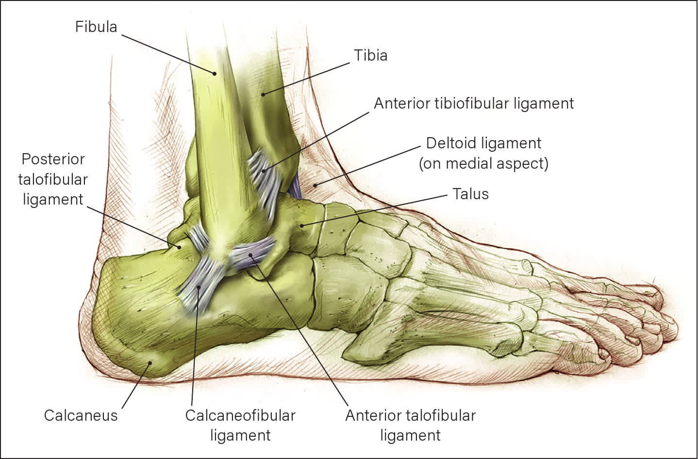
Lateral Ankle Joint Anatomy
Illustration by Christy Krames.
Reprinted with permission.
Figure 8
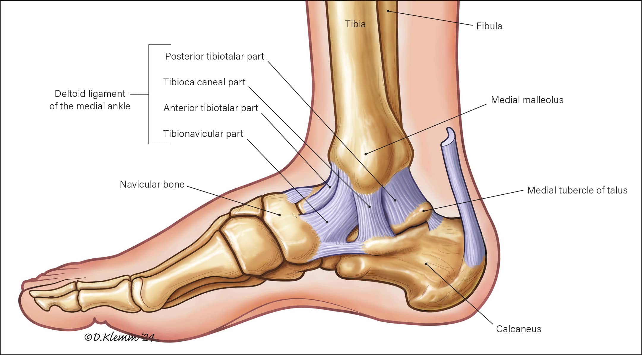
Medial Ankle Joint Anatomy
Illustration by Dave Klemm.
Figure 9
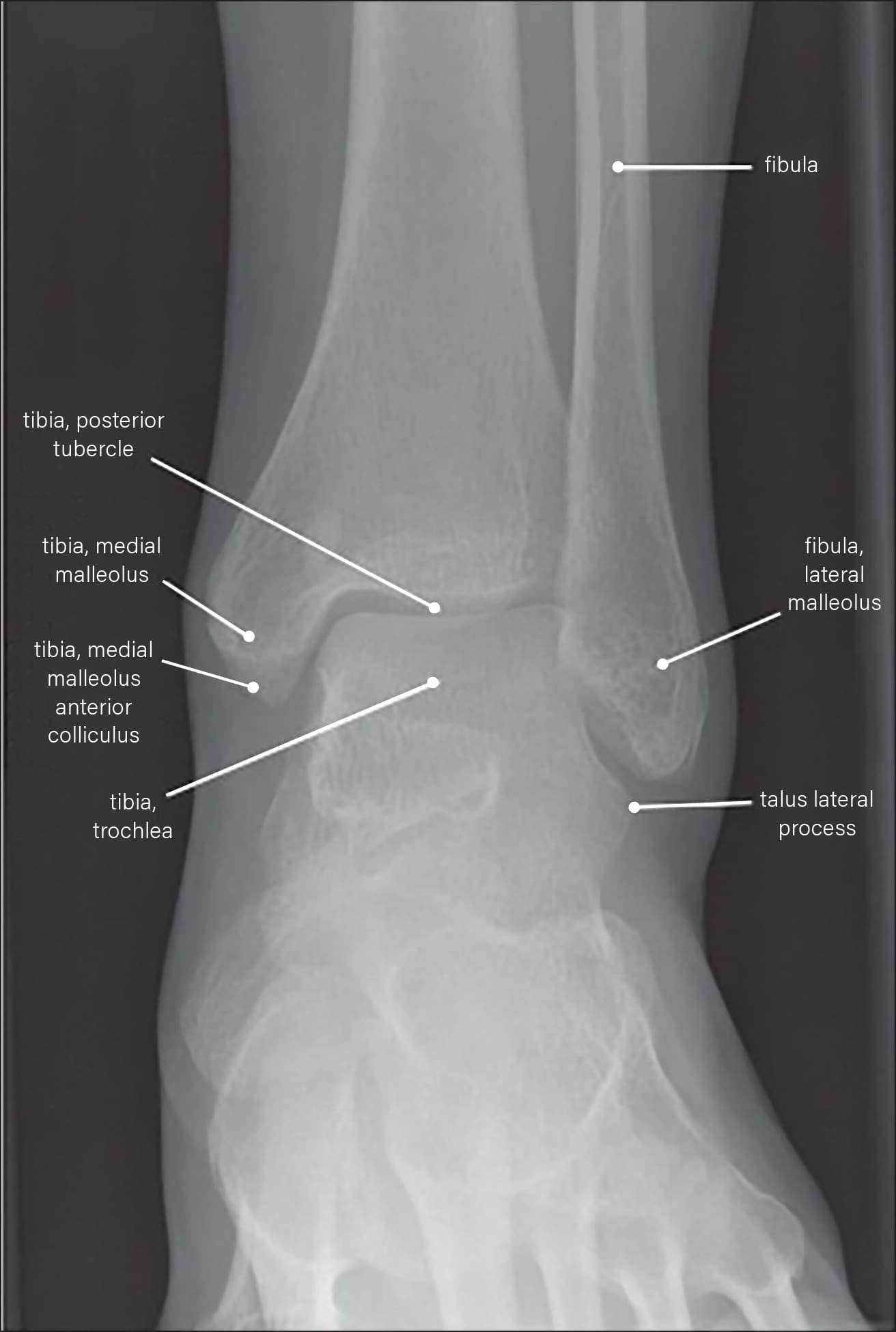
Labeled Anteroposterior Ankle X-Ray
Reprinted with permission from Omar IM. Diagnostic imaging techniques of the foot and ankle. Musculoskeletal Key. https://musculoskeletalkey.com/diagnostic-imaging-techniques-of-the-foot-and-ankle/
Figure 10
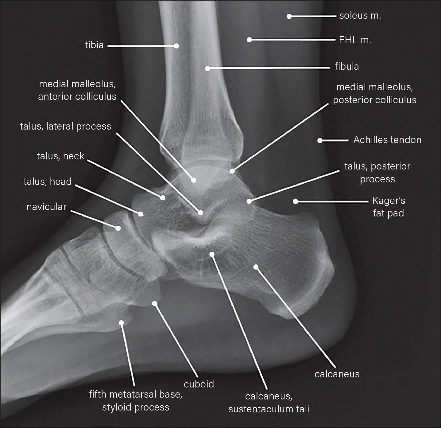
Labeled Lateral Ankle X-Ray
FHL = flexor hallucis longus.
Reprinted with permission from Omar IM. Diagnostic imaging techniques of the foot and ankle. Musculoskeletal Key. https://musculoskeletalkey.com/diagnostic-imaging-techniques-of-the-foot-and-ankle/
CLASSIFICATION OF INJURIES
Ankle injuries can result from direct trauma or a combination of force vectors.121 Injury patterns depend on the mechanism. Lateral ankle ligament injuries, sprains/tears of the ATFL, distal fibular avulsions, and physeal fractures are commonly the result of inversion or internal rotation injuries. Syndesmotic (ie, high ankle sprains), deltoid ligament, distal tibial, and proximal fibular fractures more commonly result from eversion or external rotation injuries. Ankle sprains previously were thought to be much less common, but advanced imaging has shown that ATFL sprains are more common than distal fibular physeal fractures.123
Read the full edition
Get immediate access, anytime, anywhere.
Choose a single edition, 1-year or 2-year full-access subscription.
Earn 4 CME credits for this edition.
