A lung abscess is a cavity with a well-defined wall that develops in the lung due to microbial infection. This most commonly occurs with polymicrobial aerobic and anerobic infections related to aspiration pneumonia. Lung abscess may also be related to necrotizing pneumonia from aerobic organisms (eg, Staphylococcus aureus, Pseudomonas aeruginosa), septic emboli, or bronchial obstruction (eg, tumor). Most patients respond to appropriate antimicrobial therapy. However, catheter or surgical drainage may be needed if initial therapy is ineffective or the patient has complications such as extension into the pleural space (empyema). Pleural effusion is a manifestation of various underlying pathologies with a broad differential diagnosis. Defining the etiology of pleural effusion is critical for appropriate management. Thoracentesis should be considered for all pleural effusions associated with pneumonia. Parapneumonic effusions and empyema should be treated with prompt initiation of antibiotics and drainage of infected pleural fluid.
Case 1. DB is a 36-year-old man brought to the emergency department by paramedics. He has alcohol use disorder, housing insecurity, and a 20-pack-year smoking history. He has a cough producing dark, foul-smelling sputum that has been worsening for 1 week and is now associated with midsternal and left upper back pain. DB reports weakness, dyspnea, and chills but no hemoptysis. His temperature is 102°F (38.9°C), pulse is 106 beats/min, blood pressure is 145/89 mm Hg, and pulse oximetry is 93% on room air. Auscultation reveals coarse rales in the left middle to lower lung fields and normal heart sounds. His white blood cell count is 16,000/μL (16 × 109 /L) with a normal differential. Chest radiography shows a 4-cm, left midlung cavitary lesion with a definite air-fluid level.
Lung Abscess
A lung abscess is a cavity with a well-defined wall that develops in the lung due to microbial infection.1 Abscesses are classified as primary or secondary.2 Primary lung abscesses result from direct infection of the pulmonary parenchyma in an otherwise healthy person, usually from aspiration of oral contents. Primary lung abscesses can also arise within an area of pneumonia caused by necrotizing organisms (eg, Staphylococcus aureus, Pseudomonas aeruginosa). Secondary lung abscesses occur with a predisposing condition such as bronchial obstruction (eg, foreign body, neoplasm), hematogenous spread (eg, right-sided endocarditis), or direct extension of a local infection (eg, empyema).
EPIDEMIOLOGY
The incidence of lung abscess is not well described but has markedly decreased since the introduction of effective antimicrobials. Lung abscesses are more common in patients with periodontal disease or increased occurrence of dysphagia and aspiration (eg, alcoholism).3
PATHOPHYSIOLOGY
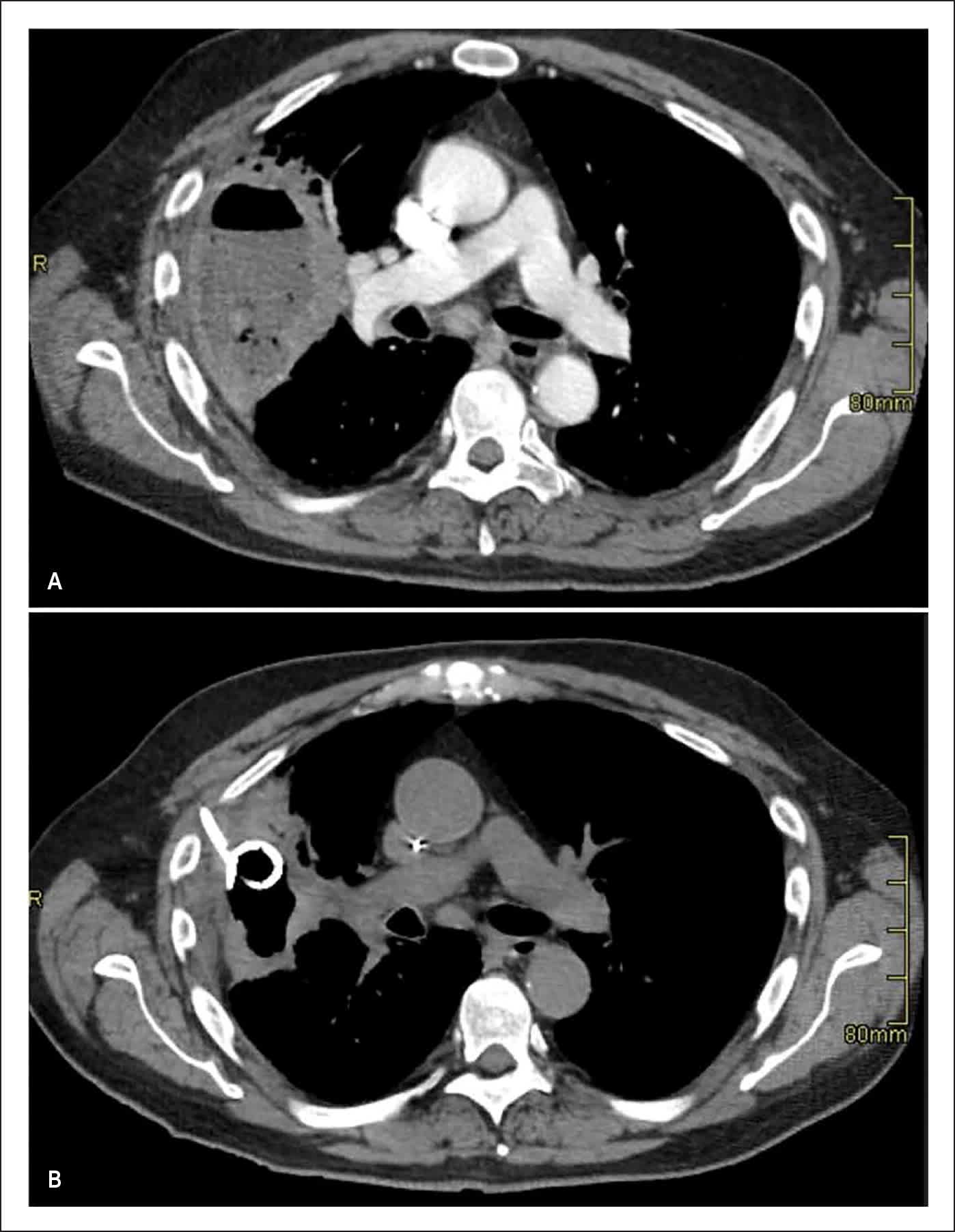
Lung abscesses occur when there are necrosis and subsequent liquefaction of lung tissue followed by removal of inflammatory debris via drainage of the bronchial tree. Communication with the bronchial tree allows for air within the cavity and inflammatory fluid drainage. Some fluid often remains, as demonstrated by the presence of an air-fluid level on imaging (Figure 1).
FIGURE 1
Computed Tomography of Lung Abscess
Images showing a large right-sided, fluid-filled lung abscess (A) before and (B) after drainage with catheter insertion.
The most common primary lung abscess is a consequence of community-acquired aspiration pneumonia, which occurs due to the microaspiration of oropharyngeal secretions.3 Infection is often polymicrobial, which can be associated with microbial synergy (the process by which the combination of microorganisms increases the pathogenicity) and leads to necrotizing pneumonia. Certain aerobic bacteria can also produce necrosis that causes a cavitating lung abscess, most commonly, S aureus, Klebsiella, and P aeruginosa, which are more likely associated with nosocomial pneumonia.2,4
Read the full edition
Get immediate access, anytime, anywhere.
Choose a single edition, 1-year or 2-year full-access subscription.
Earn 4 CME credits for this edition.