
Of the 550 million people worldwide and 37 million people in the United States with diabetes, more than 30% will develop a diabetic foot ulcer. Patients with diabetes are at increased risk of foot ulceration due to neuropathic loss of protective sensation, muscle atrophy and repetitive stress leading to foot deformities, and vascular disease. Periodic diabetic foot examinations performed by the primary care physician can identify preulcerative conditions. If present, these should prompt interventions, such as offloading for the area of concern, patient education, prescriptions for diabetic or custom footwear, or a referral to podiatry. When an ulcer is identified, classification using the SINBAD (site, ischemia, neuropathy, bacterial infection, area [ie, size], depth) system is beneficial for triage in primary care. Initial diabetic foot wound care should begin with assessment in primary care; offloading of the ulcer; treatment of infection if present; and prompt referral to specialists in wound care, infectious disease, or vascular surgery. If signs of ischemia (eg, decreased pedal pulses or blood flow) are identified, patients should be quickly referred for arterial duplex ultrasonography; if the signs of ischemia are more marked, immediate referral for vascular surgery is indicated.
Case 1. AO, a 60-year-old patient who self-identifies as mixed African and Hispanic heritage, has type 2 diabetes with neuropathy, a 35 pack-year smoking history, and a right below-knee amputation. Her chief concern today is a sore on her left plantar foot. She ambulates with use of a prosthetic and wears off-the-shelf sneakers. An ulcer is present on the plantar surface of the left midfoot. The dorsalis pedis pulse is faint but palpable, and the plantar foot is diffusely insensate. Fluctuance, discoloration, and scant purulent discharge from the wound are consistent with infection.
Epidemiology and Pathophysiology
Approximately 550 million people worldwide and 37 million people in the United States have been diagnosed with diabetes; more than 30% of those people will develop a diabetic foot ulcer during their lifetime. Among patients with diabetic ulcers, 50% will develop an infection, and 20% will undergo a lower extremity amputation.1
Diabetic foot ulcers are categorized differently than other wounds because of an etiologic triad: neuropathic loss of protective sensation, muscle atrophy and repetitive stress leading to foot deformities, and vascular disease.2 Autonomic neuropathy leads to skin changes contributing to the development of preulcerative calluses and fissures.3 These changes cause abnormal foot pressures that are not sensed by the patient. Any repetitive stress, such as walking, friction inside shoes, or shifting of weight, can cause blistering or hemorrhage under calluses.1 To compound this, patients may have localized ischemia due to ill-fitting shoes constantly pressing against insensate tissue, or direct trauma from an unfelt impact or foreign body.1,4 Figure 1 illustrates diabetic foot ulcerations. Most foot ulcers associated with diabetes have a mixed etiology (Table 12,3,5–7).
Figure 1
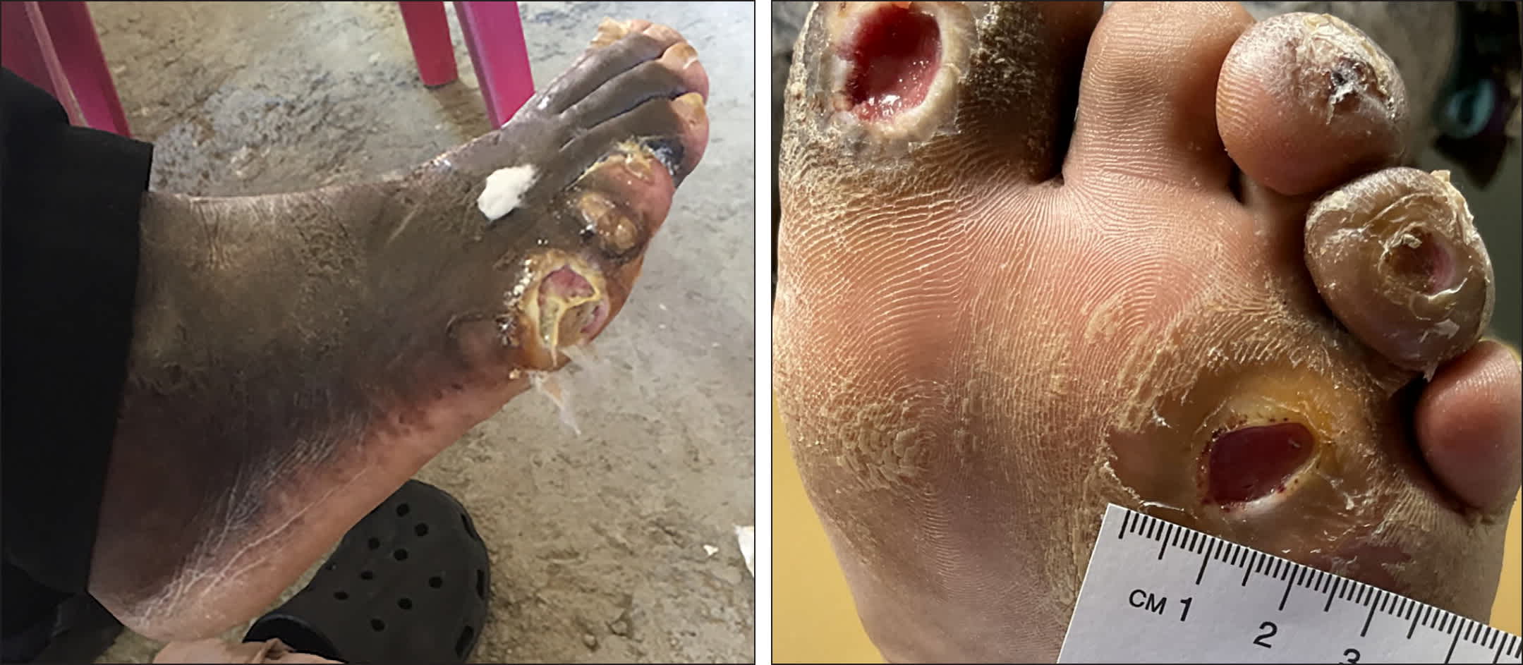
Diabetic Foot Ulcers
Examples of diabetic foot ulcers secondary to diabetic foot triad.
Table 1 Etiologies of Foot Ulcers
| Type | Etiology | Differentiating factors |
|---|---|---|
| Diabetic foot ulcers3,5 | Loss of protective sensation, muscle atrophy and repetitive stress leading to foot deformities, and vascular disease | History of poor glycemic control; may have preulcerative lesions or foot deformities |
| Pressure ulcers2,6,7 | Compression of tissue (or the artery leading to the tissue) causes local ischemia, and shear or friction forces pull and damage the tissue over bony prominences | Associated with an area of pressure due to an ill-fitting shoe, footrests, or hospital bed; patients may be nonambulatory or have difficulty voluntarily repositioning |
| Arterial ulcers3,7 | Reduced arterial blood flow secondary to atherosclerosis, diabetic microvascular disease, vasculitis, or arterial emboli | History of intermittent claudication, worsening pain when leg is elevated due to draining blood from the wound area, even in the presence of neuropathy; decreased dorsalis pedis pulse with or without decreased posterior tibialis pulse; wound areas have sharply defined edges (ie, punched out), are painful to touch, deep, and often covered with black necrotic tissue (eschar) |
Information from references 2, 3, and 5-7.
Read the full edition
Get immediate access, anytime, anywhere.
Choose a single edition, 1-year or 2-year full-access subscription.
Earn 4 CME credits for this edition.