
Primary care physicians often serve as the initial point of contact for patients presenting with potential fractures. Initial evaluation with plain radiography or ultrasonography can confirm the diagnosis. If clinical suspicion remains high despite negative imaging, the joint should be immobilized with protected weight-bearing. Then, reassessment should include repeat radiography in 10 to 14 days or advanced imaging with computed tomography or magnetic resonance imaging without intravenous contrast based on location and risk. For fractures that can be managed nonoperatively, the joint should be immobilized with a brace or cast, with short-interval follow-up and repeat radiography. Common indications for referral to an orthopedic surgeon include fractures that are open, displaced, intra-articular, unstable, fracture-dislocations, and those with neurovascular compromise. Fractures with nonunion or malunion should be managed surgically. Rehabilitation is supported by some evidence but requires further research to define optimal protocols for specific fracture types. Special populations, including older adults and athletes, require tailored care plans. Multidisciplinary care has been shown to improve outcomes in patients with complex fractures and in older adults. Racial and ethnic disparities in morbidity and mortality rates associated with fractures highlight the need for improved access to quality care.
Case 1. JM is a 32-year-old woman with a distal radius fracture from a bicycle crash. She presents to the clinic wearing an arm splint she received 3 days ago at an urgent care center. She is asking about next steps in management, potential complications, and when she can return to competitive cycling.
Common Fractures in Primary Care
Primary care physicians frequently serve as the initial point of contact for patients with fractures, necessitating a strong understanding of initial assessment and management. A variety of simple fractures can be managed by family physicians. Knowing when to refer to specialists is a vital aspect of management.
In adults, common fracture sites in the upper extremities include the proximal humerus, distal radius, metacarpals, and phalanges; in the lower extremities, common fracture sites include the femur, tibia, and fibula.1–3 Vertebral compression fractures are most commonly seen in older adults and are associated with low bone mineral density.4
Diagnosis
HISTORY
A thorough patient history that includes the mechanism of injury will provide clues as to the likely fracture pattern and associated soft tissue damage (Figure 11,5–9). Patient demographics, including age, physical activity level, comorbid conditions (eg, osteoporosis, diabetes), and current medications (eg, anticoagulants) significantly influence treatment planning and may help predict potential complications.10 Most patients with a fracture report symptoms of focal pain and swelling, and those with a lower extremity fracture may report difficulty with weight-bearing.11
Figure 1
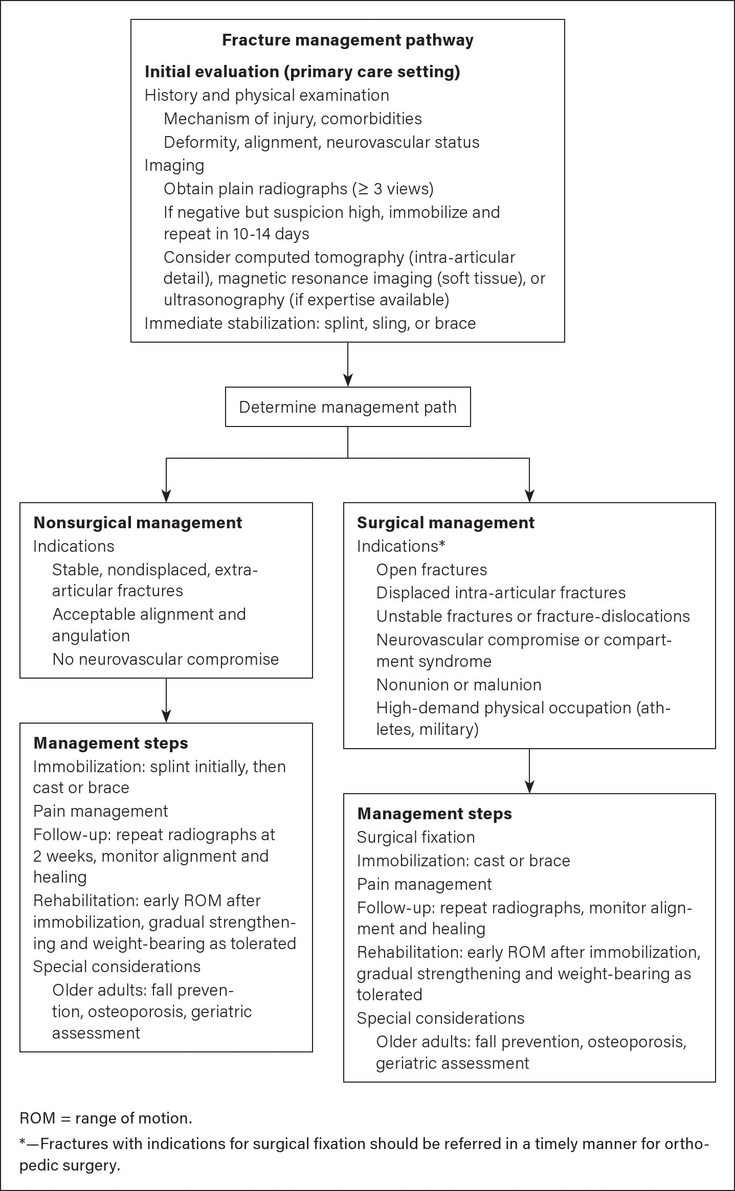
Clinical Decision-Making Guidance for Fracture Assessment and Management in Adults in the Primary Care Setting
PHYSICAL EXAMINATION
In a patient with a known or suspected fracture, a comprehensive physical examination can identify associated soft tissue injuries, neurovascular compromise, and the extent of bony deformity. Careful inspection will determine whether the fracture is open or closed, as well as the extent of deformity. Rotational and angular deformities such as those of the hand should be assessed by asking the patient to make a fist to evaluate alignment of the metacarpophalangeal and phalangeal joints.2 Palpation of osseous structures and soft tissues aids in determining maximal point of tenderness.
Read the full edition
Get immediate access, anytime, anywhere.
Choose a single edition, 1-year or 2-year full-access subscription.
Earn 4 CME credits for this edition.