
Lower extremity edema results from an imbalance between capillary hydrostatic pressure, oncotic pressure, and lymphatic drainage, leading to fluid accumulation in the interstitial space. Most cases are bilateral, typically due to systemic causes of volume overload such as heart failure, cirrhosis, or kidney dysfunction. Unilateral edema is more often due to localized pathology, including deep venous thrombosis, cellulitis, or trauma. Edema is a common presenting symptom and may be associated with pain, reduced mobility, and functional impairment. Initial evaluation should include a history and physical examination with attention to risk factors, symptom duration, laterality, precipitating factors, and associated signs and symptoms. Laboratory tests and diagnostic imaging are selected based on clinical suspicion, in some cases aided by tools such as the Wells criteria for deep venous thrombosis. Management targets the underlying etiology. Edema due to systemic causes often improves with disease-specific therapy. Edema due to chronic venous insufficiency and lymphedema respond to compression therapy. Lipedema is the painful localized accumulation of subcutaneous adipose tissue and treatment is focused on symptom management.
Case 4. BC is a 59-year-old with a history of hypertension, hyperlipidemia, type 2 diabetes, moderate obesity, and remote tobacco use. He presents with a concern of worsening bilateral lower extremity swelling. He states that the nature of his work involves standing for most of the day, and by the end of the day his legs are swollen. On most mornings, the swelling has completely subsided, but over the past few weeks it has been persistent. He denies any recent chest pain, shortness of breath, or palpitations, but believes that he has recently gained a few pounds.
Risk Factors and Impact
Edema is the excess accumulation of fluid in the interstitial space that is caused by an imbalance in capillary hemodynamics. Up to 20% of adults older than 50 years have peripheral edema of the legs.1 Older age, female sex, obesity, diabetes, hypertension, and inactivity are all associated with an increased incidence of lower extremity edema.1
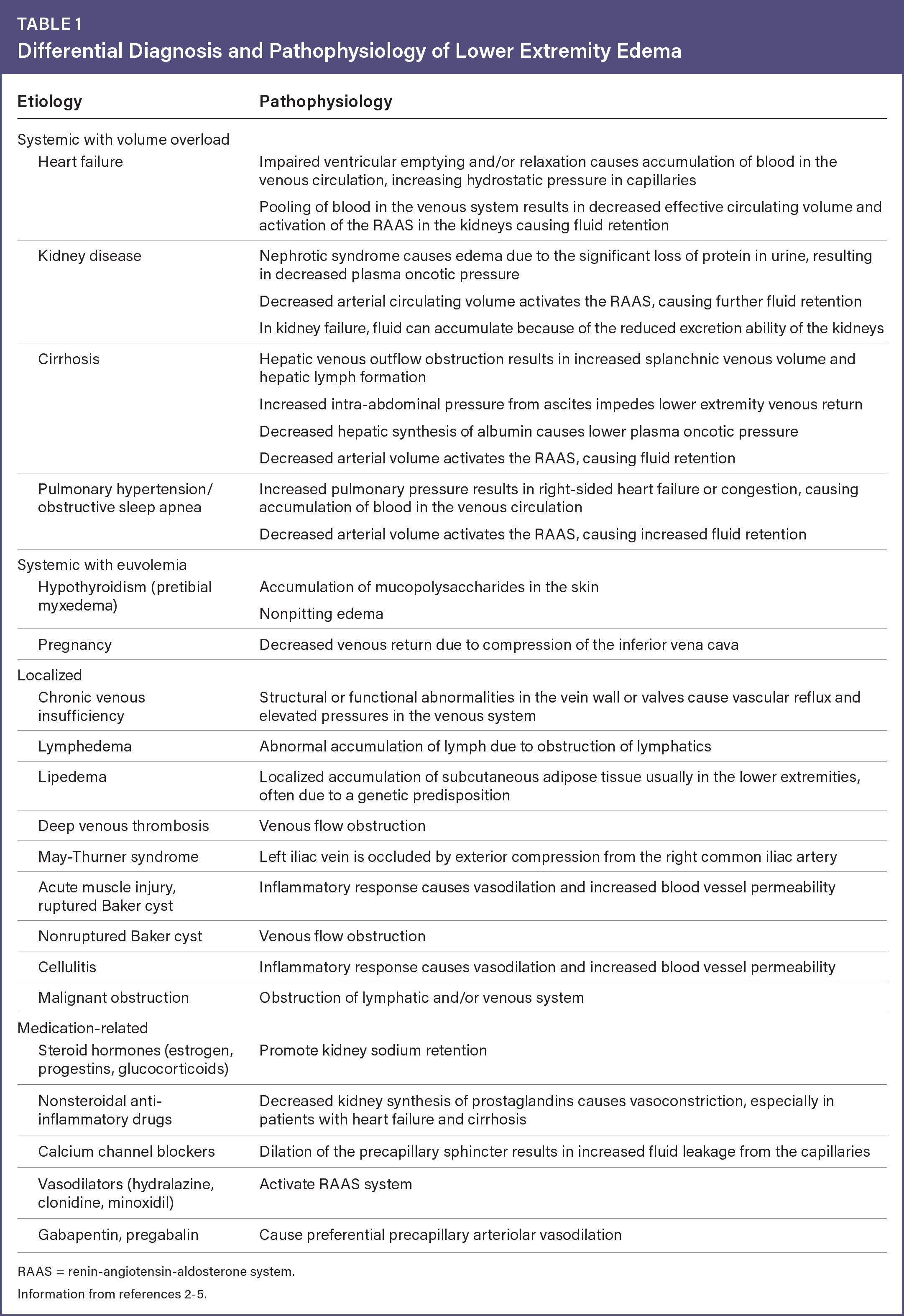
Peripheral edema has a significant impact on quality of life, with 65% of affected patients reporting at least mild pain most of the time.1 The condition may also lead to reduced mobility and functional impairment. The differential diagnosis of lower extremity edema and pathophysiology of each cause are listed in Table 1.2–5
Table 1 Differential Diagnosis and Pathophysiology of Lower Extremity Edema
| Etiology | Pathophysiology |
|---|---|
| Systemic with volume overload | |
| Heart failure | Impaired ventricular emptying and/or relaxation causes accumulation of blood in the venous circulation, increasing hydrostatic pressure in capillaries Pooling of blood in the venous system results in decreased effective circulating volume and activation of the RAAS in the kidneys causing fluid retention |
| Kidney disease | Nephrotic syndrome causes edema due to the significant loss of protein in urine, resulting in decreased plasma oncotic pressure Decreased arterial circulating volume activates the RAAS, causing further fluid retention In kidney failure, fluid can accumulate because of the reduced excretion ability of the kidneys |
| Cirrhosis | Hepatic venous outflow obstruction results in increased splanchnic venous volume and hepatic lymph formation Increased intra-abdominal pressure from ascites impedes lower extremity venous return Decreased hepatic synthesis of albumin causes lower plasma oncotic pressure Decreased arterial volume activates the RAAS, causing fluid retention |
| Pulmonary hypertension/obstructive sleep apnea | Increased pulmonary pressure results in right-sided heart failure or congestion, causing accumulation of blood in the venous circulation Decreased arterial volume activates the RAAS, causing increased fluid retention |
| Systemic with euvolemia | |
| Hypothyroidism (pretibial myxedema) | Accumulation of mucopolysaccharides in the skin Nonpitting edema |
| Pregnancy | Decreased venous return due to compression of the inferior vena cava |
| Localized | |
| Chronic venous insufficiency | Structural or functional abnormalities in the vein wall or valves cause vascular reflux and elevated pressures in the venous system |
| Lymphedema | Abnormal accumulation of lymph due to obstruction of lymphatics |
| Lipedema | Localized accumulation of subcutaneous adipose tissue usually in the lower extremities, often due to a genetic predisposition |
| Deep venous thrombosis | Venous flow obstruction |
| May-Thurner syndrome | Left iliac vein is occluded by exterior compression from the right common iliac artery |
| Acute muscle injury, ruptured Baker cyst | Inflammatory response causes vasodilation and increased blood vessel permeability |
| Nonruptured Baker cyst | Venous flow obstruction |
| Cellulitis | Inflammatory response causes vasodilation and increased blood vessel permeability |
| Malignant obstruction | Obstruction of lymphatic and/or venous system |
| Medication-related | |
| Steroid hormones (estrogen, progestins, glucocorticoids) | Promote kidney sodium retention |
| Nonsteroidal anti-inflammatory drugs | Decreased kidney synthesis of prostaglandins causes vasoconstriction, especially in patients with heart failure and cirrhosis |
| Calcium channel blockers | Dilation of the precapillary sphincter results in increased fluid leakage from the capillaries |
| Vasodilators (hydralazine, clonidine, minoxidil) | Activate RAAS system |
| Gabapentin, pregabalin | Cause preferential precapillary arteriolar vasodilation |
RAAS = renin-angiotensin-aldosterone system.
Read the full edition
Get immediate access, anytime, anywhere.
Choose a single edition, 1-year or 2-year full-access subscription.
Earn 4 CME credits for this edition.