Syncope is an abrupt, transient, and complete loss of consciousness associated with an inability to maintain postural tone, followed by rapid and spontaneous recovery. Syncope is caused by temporary cerebral hypoperfusion. Presyncope describes symptoms such as lightheadedness and vision changes that may or may not precede syncope. Syncope can be classified by its mechanism: reflex (neurally mediated), orthostatic hypotension, or cardiac. Initial evaluation of loss of consciousness should include a careful history and detailed physical examination with orthostatic vital signs and 12-lead electrocardiography. The history should evaluate whether the episode was a true loss of consciousness and clarify whether it has a syncopal or nonsyncopal etiology. Although there is no evidence-based standard for the diagnosis of syncope, consensus suggests diagnostic criteria concordant with the three syncope mechanisms. The treatment of syncope is specific to its mechanism. Multiple risk stratification tools exist; however, these do not outperform clinician judgment in predicting serious outcomes of syncope in the short term. When evaluating and treating syncope, additional consideration should be given to special populations including children and adolescents, older adults, athletes, and patients with postural orthostatic tachycardia syndrome.
Case 3. LH is an 18-year-old, otherwise healthy college student and vocal performance major who presents after several episodes of passing out at school. She states that these episodes have occurred randomly. She suddenly feels lightheaded, even if she has eaten recently. Before the episodes of passing out, or feeling like she will pass out, she has some nausea and blurry vision, but has not had headache, chest pain, or any neurologic signs or symptoms.
Definitions and Pathophysiology
Syncope is an abrupt, transient, and complete loss of consciousness associated with an inability to maintain postural tone, followed by rapid and spontaneous recovery. Syncope excludes other causes of loss of consciousness such as head trauma, seizures, metabolic conditions, and intoxication. The specific characteristics of syncope include a short duration of loss of responsiveness and abnormal motor control with amnesia during the event. Presyncope, or near syncope, describes the symptoms preceding syncope (eg, lightheadedness and vision changes such as tunnel vision or graying out), which may or may not progress to syncope.
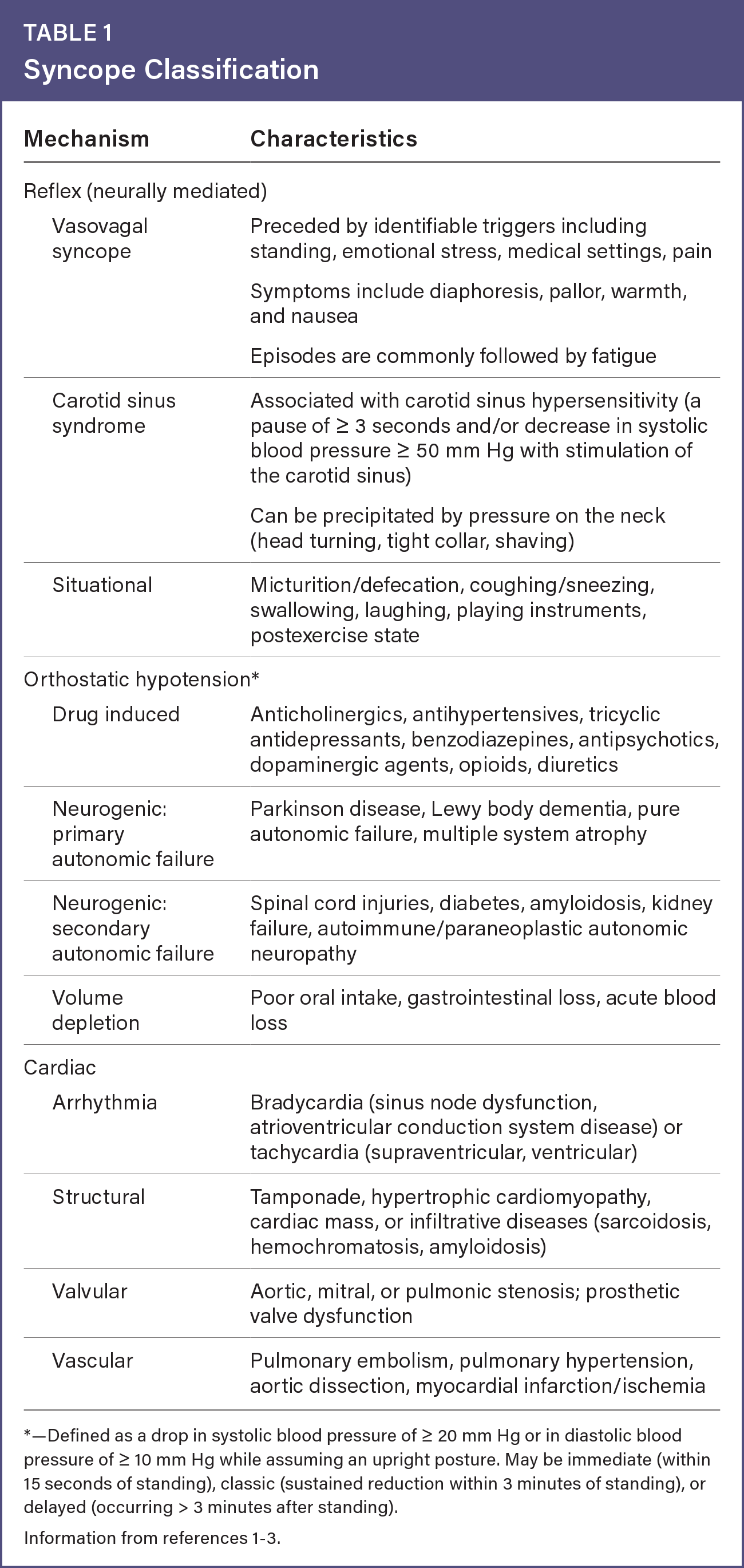
The pathophysiology of syncope is presumed to be related to cerebral hypoperfusion, which can occur after loss of cerebral blood flow for as little as 6 to 8 seconds. Variable degrees of decreased peripheral resistance (vasodilation) and decreased cardiac output, separately or in tandem, can cause cerebral hypoperfusion. Syncope can be classified according to three overarching mechanisms—reflex (neurally mediated), orthostatic hypotension, or cardiac—each having subtypes with specific characteristics (Table 11–3). Nonsyncopal transient loss of consciousness can have cardiac, metabolic, or neurologic etiologies, and psychogenic pseudosyncope represents an episode of apparent syncope with no true loss of consciousness.1,2
Table 1 Syncope Classification
| Mechanism | Characteristics |
|---|---|
| Reflex (neurally mediated) | |
| Vasovagal syncope | Preceded by identifiable triggers including standing, emotional stress, medical settings, pain Symptoms include diaphoresis, pallor, warmth, and nausea Episodes are commonly followed by fatigue |
| Carotid sinus syndrome | Associated with carotid sinus hypersensitivity (a pause of ≥ 3 seconds and/or decrease in systolic blood pressure ≥ 50 mm Hg with stimulation of the carotid sinus) Can be precipitated by pressure on the neck (head turning, tight collar, shaving) |
| Situational | Micturition/defecation, coughing/sneezing, swallowing, laughing, playing instruments, postexercise state |
| Orthostatic hypotension* | |
| Drug induced | Anticholinergics, antihypertensives, tricyclic antidepressants, benzodiazepines, antipsychotics, dopaminergic agents, opioids, diuretics |
| Neurogenic: primary autonomic failure | Parkinson disease, Lewy body dementia, pure autonomic failure, multiple system atrophy |
| Neurogenic: secondary autonomic failure | Spinal cord injuries, diabetes, amyloidosis, kidney failure, autoimmune/paraneoplastic autonomic neuropathy |
| Volume depletion | Poor oral intake, gastrointestinal loss, acute blood loss |
| Cardiac | |
| Arrhythmia | Bradycardia (sinus node dysfunction, atrioventricular conduction system disease) or tachycardia (supraventricular, ventricular) |
| Structural | Tamponade, hypertrophic cardiomyopathy, cardiac mass, or infiltrative diseases (sarcoidosis, hemochromatosis, amyloidosis) |
| Valvular | Aortic, mitral, or pulmonic stenosis; prosthetic valve dysfunction |
| Vascular | Pulmonary embolism, pulmonary hypertension, aortic dissection, myocardial infarction/ischemia |
*—Defined as a drop in systolic blood pressure of ≥ 20 mm Hg or in diastolic blood pressure of ≥ 10 mm Hg while assuming an upright posture. May be immediate (within 15 seconds of standing), classic (sustained reduction within 3 minutes of standing), or delayed (occurring > 3 minutes after standing).
Read the full edition
Get immediate access, anytime, anywhere.
Choose a single edition, 1-year or 2-year full-access subscription.
Earn 4 CME credits for this edition.