

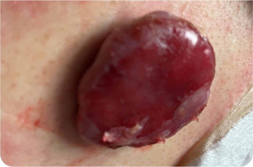
A 26-year-old man presented to the emergency department after having a five-minute tonic-clonic seizure. He was postictal for less than one hour and then returned to his baseline. Over the previous two days, he had experienced a mild diffuse headache and an intermittent blind spot in the center of his vision. For the previous three weeks, he had noticed left upper extremity weakness and dexterity issues in his left hand occurring after intense sessions of upper extremity weight lifting. He also reported having a lesion on his back that had been slowly growing for more than one year and occasionally bled. Otherwise, the patient appeared healthy. He had no history of recent travel.
Physical examination confirmed left upper extremity weakness and impaired fine motor skills of the fingers. The patient had normal heart sounds. The remainder of the neurologic examination was unremarkable. Magnetic resonance imaging (MRI) of the brain was performed (Figure 1). A pedunculated lesion measuring 4.0 × 3.5 × 1.5 cm (Figure 2) was found on his left upper back.
FIGURE 1
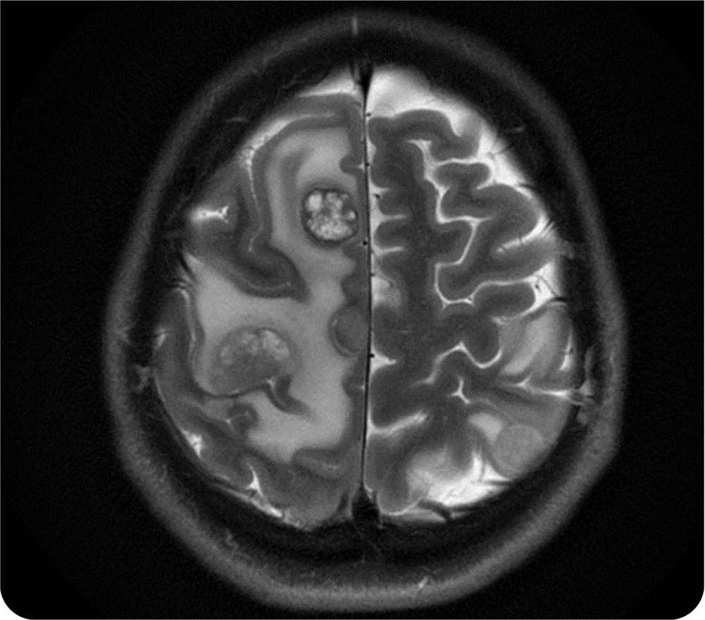
FIGURE 2
Question
Based on the patient's history and physical examination findings, which one of the following is the most likely diagnosis?
- A. Brain abscess.
- B. Glioma with high-grade features.
- C. Hemangioma.
- D. Nodular melanoma.
Discussion
The answer is D: nodular melanoma, leading to brain metastasis. Nodular melanoma is the second most common type of melanoma, and it is the most aggressive form. The lesion can be pedunculated, polypoid, or an elevated plaque with ulcerations that may bleed intermittently. The primary lesion is commonly located on the trunk or limbs.1
Melanoma is the most lethal cutaneous malignancy; the five-year survival rate is 30% for patients with stage IV disease. However, early detection is associated with a 99.4% survival rate for patients with stages I and II disease.2 Risk factors for melanoma are sun exposure, indoor tanning, immunosuppression, personal history of nevi, family history of melanoma, and obesity.3
Any suspicious lesions should be completely excised, and a 1- to 3-mm border of healthy skin should also be excised around each lesion. Subsequent wide excision surgical treatment is based on biopsy results and disease staging. Treatment options for melanoma with large or symptomatic brain metastasis are limited to stereotactic radiosurgery, surgical resection with postoperative radiation therapy, or whole-brain radiation therapy. Approximately 50% of melanomas are associated with BRAF mutation (V600 mutation is the most common).4 Immunotherapy using ipilimumab (Yervoy) with nivolumab (Opdivo) may be effective for advanced disease in patients with the BRAF V600 mutation.
Brain abscesses can present with unilateral or progressive diffuse headache (69% of patients), fever (53%), focal neurologic deficits (48%), and seizures (25% have seizure as the first manifestation). The classic triad of headache, fever, and focal neurologic deficit occurs in less than 20% of patients.5 MRI is preferred for diagnosis and typically demonstrates a ring-enhancing lesion with central necrosis and vasogenic edema.
Gliomas with high-grade features typically develop over days to weeks, often with headache, seizures, and focal neurologic symptoms. MRI demonstrates an isolated ring-enhancing lesion with necrosis.6 Multiple lesions in the brain are uncommon; they are more typical of metastatic disease than primary brain malignancy.
Read the full article
Get immediate access, anytime, anywhere.
Choose a single article, issue, or full-access subscription.
Earn up to 14 CME credits per issue.
