

CCTA to Evaluate for Coronary Artery Stenosis in Intermediate-Risk Patients With Stable Chest Pain

Allison Karlan Kaplan, MD
Nicholas Whitbeck, OMS4

American Family Physician. 2024;110(3):298-300.

Author disclosure: No relevant financial relationships.

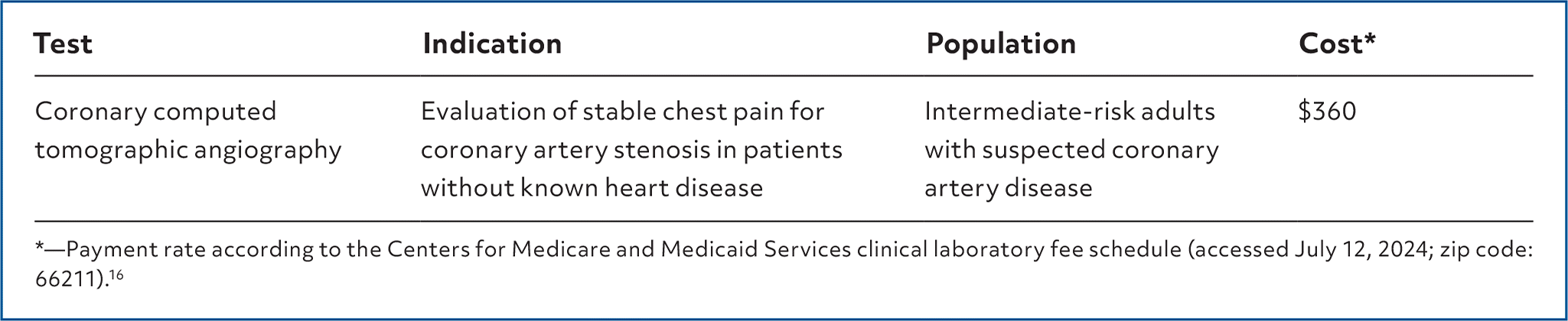
TestIndicationPopulationCost*
Coronary computed tomographic angiographyEvaluation of stable chest pain for coronary artery stenosis in patients without known heart diseaseIntermediate-risk adults with suspected coronary artery disease$360

*—Payment rate according to the Centers for Medicare and Medicaid Services clinical laboratory fee schedule (accessed July 12, 2024; zip code: 66211).16

Coronary computed tomographic angiography (CCTA) is a noninvasive radiology test used to evaluate for coronary artery stenosis by detecting high-risk atherosclerotic plaques and their volume and location.1 In 2021, the American College of Cardiology/American Heart Association Joint Committee recommended CCTA over invasive coronary angiography (ICA) or functional imaging for intermediate-risk patients with stable chest pain and no known coronary artery disease (CAD) to rule out coronary stenosis.2

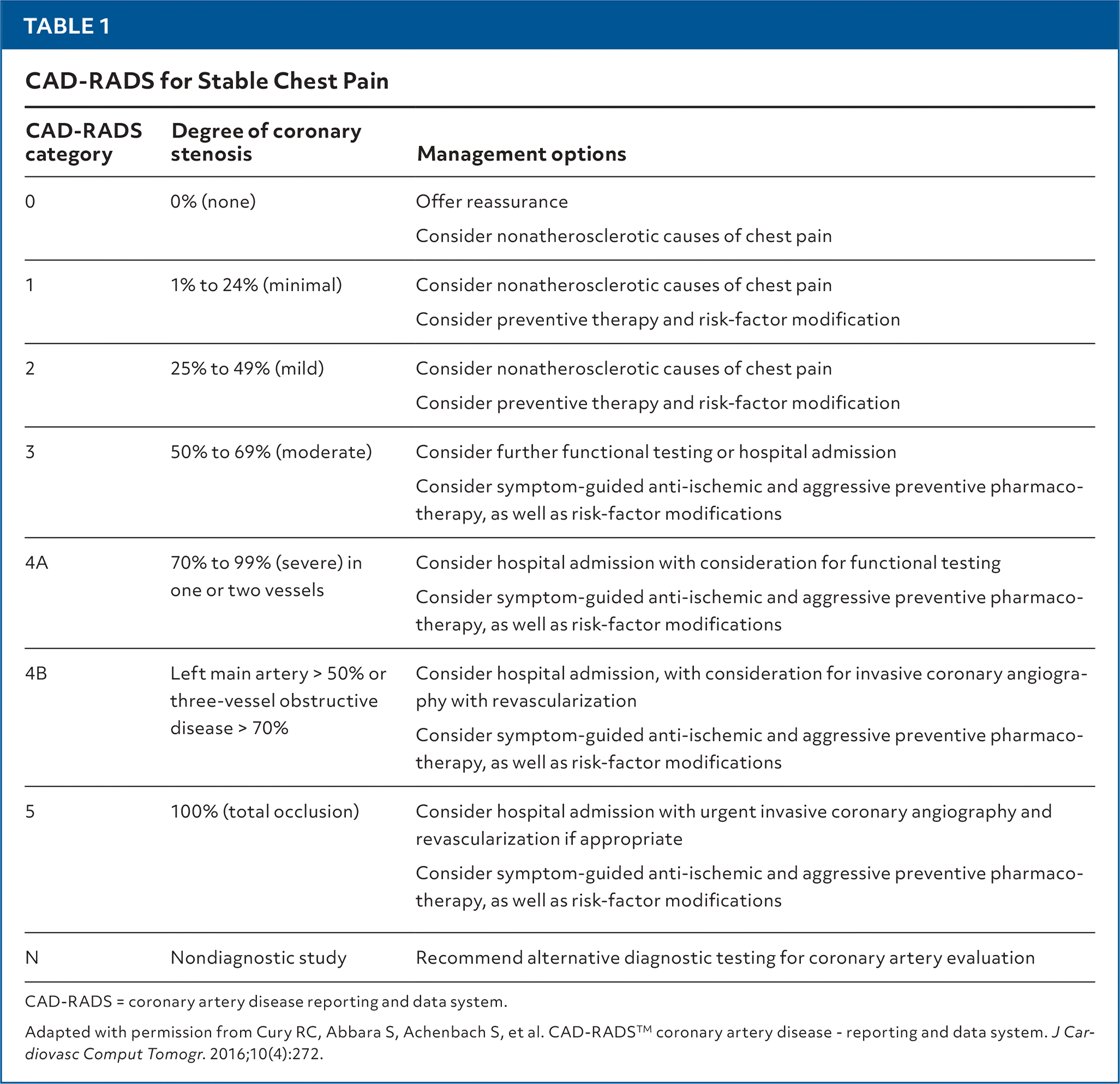
Intermediate risk can be defined using different clinical decision pathways such as the HEART (history, electrocardiography, age, risk factors, and troponin) score for major cardiac events and the Thrombolysis in Myocardial Infarction score. The classification system groups CCTA findings from 0 to 5 based on the degree of coronary stenosis and location and recommends management options (Table 1).3

TABLE 1. CAD-RADS for Stable Chest Pain
CAD-RADS categoryDegree of coronary stenosisManagement options
00% (none)Offer reassurance
Consider nonatherosclerotic causes of chest pain
11% to 24% (minimal)Consider nonatherosclerotic causes of chest pain
Consider preventive therapy and risk-factor modification
225% to 49% (mild)Consider nonatherosclerotic causes of chest pain
Consider preventive therapy and risk-factor modification
350% to 69% (moderate)Consider further functional testing or hospital admission
Consider symptom-guided anti-ischemic and aggressive preventive pharmacotherapy, as well as risk-factor modifications
4A70% to 99% (severe) in one or two vesselsConsider hospital admission with consideration for functional testing
Consider symptom-guided anti-ischemic and aggressive preventive pharmacotherapy, as well as risk-factor modifications
4BLeft main artery > 50% or three-vessel obstructive disease > 70%Consider hospital admission, with consideration for invasive coronary angiography with revascularization
Consider symptom-guided anti-ischemic and aggressive preventive pharmacotherapy, as well as risk-factor modifications
5100% (total occlusion)Consider hospital admission with urgent invasive coronary angiography and revascularization if appropriate
Consider symptom-guided anti-ischemic and aggressive preventive pharmacotherapy, as well as risk-factor modifications
NNondiagnostic studyRecommend alternative diagnostic testing for coronary artery evaluation

CAD-RADS = coronary artery disease reporting and data system.

Adapted with permission from Cury RC, Abbara S, Achenbach S, et al. CAD-RADSTM coronary artery disease - reporting and data system. J Cardiovasc Comput Tomogr. 2016;10(4):272.

ACCURACY

A study comparing CCTA with ICA as the reference standard in intermediate- to high-risk patients (n = 64 patients; 827 segments) showed that CCTA had a diagnostic accuracy of 96% for detecting 50% or greater luminal stenosis (sensitivity = 87%; specificity = 97%; positive likelihood ratio = 29; negative likelihood ratio = 0.13). In segments with 70% or greater luminal stenosis, diagnostic accuracy was 99% (sensitivity = 96%; specificity = 99%; positive likelihood ratio = 96; negative likelihood ratio = 0.04). For coronary artery segments analyzed in this study, 1% were considered uninterpretable because of motion artifacts, extensive calcifications, small vessel diameter, or blooming artifact from a stent.4

ALLISON KARLAN KAPLAN, MD, FAAFP, Mountain Vista Medical Center Family Medicine Residency, Mesa, Arizona.

NICHOLAS WHITBECK, OMS4, Midwestern University, Glendale, Arizona.

Author disclosure: No relevant financial relationships.

  1. 1.Ferencik M, Mayrhofer T, Bittner DO, et al. Use of high-risk coronary atherosclerotic plaque detection for risk stratification of patients with stable chest pain: a secondary analysis of the PROMISE randomized clinical trial. JAMA Cardiol. 2018;3(2):144-152.
  2. 2.Gulati M, Levy PD, Mukherjee D, et al. 2021 AHA/ACC/ASE/Chest/SAEM/SCCT/SCMR guideline for the evaluation and diagnosis of chest pain: a report of the American College of Cardiology/American Heart Association Joint Committee on clinical practice guidelines [published corrections appear in Circulation. 2021;144(22):e455 and Circulation. 2023;148(24):e281]. Circulation. 2021;144(22):e368-e454.
  3. 3.Cury RC, Abbara S, Achenbach S, et al. CAD-RADSTM coronary artery disease - reporting and data system. J Cardiovasc Comput Tomogr. 2016;10(4):269-281.
  4. 4.de Graaf FR, Schuijf JD, van Velzen JE, et al. Diagnostic accuracy of 320-row multidetector computed tomography coronary angiography in the non-invasive evaluation of significant coronary artery disease. Eur Heart J. 2010;31(15):1908-1915.
  5. 5.Nielsen LH, Ortner N, Nørgaard BL, et al. The diagnostic accuracy and outcomes after coronary computed tomography angiography vs. conventional functional testing in patients with stable angina pectoris: a systematic review and meta-analysis. Eur Heart J Cardiovasc Imaging. 2014;15(9):961-971.
  6. 6.Aziz MU, Singh S. Computed tomography of coronary artery atherosclerosis: a review. J Med Imaging Radiat Sci. 2021;52(3S):S19-S39.
  7. 7.Schönenberger E, Schnapauff D, Teige F, et al. Patient acceptance of noninvasive and invasive coronary angiography. PLoS One. 2007;2(2):e246.
  8. 8.Al-Abdouh A, Mhanna M, Jabri A, et al. Meta-analysis of coronary computed tomography versus invasive coronary angiography in stable chest pain. Am J Cardiol. 2022;183:153-154.
  9. 9.Newby DE, Adamson PD, Berry C, et al.; SCOT-HEART Investigators. Coronary CT angiography and 5-year risk of myocardial infarction. N Engl J Med. 2018;379(10):924-933.
  10. 10.Patel MR, Peterson ED, Dai D, et al. Low diagnostic yield of elective coronary angiography [published correction appears in N Engl J Med. 2010;363(5):498]. N Engl J Med. 2010;362(10):886-895.
  11. 11.Maurovich-Horvat P, Bosserdt M, Kofoed KF, et al.; DISCHARGE Trial Group. CT or invasive coronary angiography in stable chest pain. N Engl J Med. 2022;386(17):1591-1602.
  12. 12.Burch RA, Siddiqui TA, Tou LC, et al. The cost effectiveness of coronary CT angiography and the effective utilization of CT-fractional flow reserve in the diagnosis of coronary artery disease. J Cardiovasc Dev Dis. 2023;10(1):25.
  13. 13.Budoff MJ, Dowe D, Jollis JG, et al. Diagnostic performance of 64-multidetector row coronary computed tomographic angiography for evaluation of coronary artery stenosis in individuals without known coronary artery disease: results from the prospective multicenter ACCURACY (Assessment by Coronary Computed Tomographic Angiography of Individuals Undergoing Invasive Coronary Angiography) trial. J Am Coll Cardiol. 2008;52(21):1724-1732.
  14. 14.Sun Z, Almutairi AMD. Diagnostic accuracy of 64 multislice CT angiography in the assessment of coronary in-stent restenosis: a meta-analysis. Eur J Radiol. 2010;73(2):266-273.
  15. 15.MDsave. CT angiography. Accessed May 12, 2024. https://www.mdsave.com/procedures/ct-angiography/d786ffc9
  16. 16.Centers for Medicare and Medicaid Services. License for use of current procedural terminology, fourth edition (“CPT”). Accessed May 12, 2024. https://www.cms.gov/medicare/physician-fee-schedule/search

This series is coordinated by Natasha Pyzocha, DO, contributing editor.

A collection of Diagnostic Tests published in AFP is available at https://www.aafp.org/afp/diagnostic.

Copyright © 2026 by the American Academy of Family Physicians.

This content is owned by the AAFP. A person viewing it online may make one printout of the material and may use that printout only for his or her personal, non-commercial reference. This material may not otherwise be downloaded, copied, printed, stored, transmitted or reproduced in any medium, whether now known or later invented, except as authorized in writing by the AAFP. See permissions for copyright questions and/or permission requests.
