

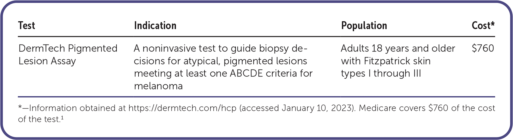
| Test | Indication | Population | Cost* |
|---|---|---|---|
| DermTech Pigmented Lesion Assay | A noninvasive test to guide biopsy decisions for atypical, pigmented lesions meeting at least one ABCDE criteria for melanoma | Adults 18 years and older with Fitzpatrick skin types I through III | $760 |
*—Information obtained at https://dermtech.com/hcp (accessed January 10, 2023). Medicare covers $760 of the cost of the test.1
The DermTech Pigmented Lesion Assay (PLA) is a noninvasive test used for the detection of melanoma. Adhesive stickers are used to collect skin cells from the entire surface of suspicious melanocytic lesions that have one or more ABCDE criteria for melanoma. These stickers are analyzed for genomic markers associated with melanoma: RNA (LINC00518 and PRAME) and optional add-on DNA (TERT). When results are positive for one or more genomic markers, a biopsy is recommended. If results are negative for genomic markers, biopsy deferral and continued monitoring of the lesion are recommended.1 The PLA has not been approved by the U.S. Food and Drug Administration.1
Accuracy
An initial validation study including 398 samples compared results from the PLA with a consensus histopathologic assessment from three dermatopathologists. Among the 398 samples (87 melanomas and 311 nonmelanomas), the PLA demonstrated a sensitivity of 91%, specificity of 69%, positive likelihood ratio of 2.94, and negative likelihood ratio of 0.13.2
For diagnostic accuracy, a pooled analysis from a systematic review of five studies (n = 2,043) comparing PLA with histopathologic examination found a sensitivity of 86.9%, specificity of 82.4%, positive likelihood ratio of 4.94, and negative likelihood ratio of 0.16.3 However, the quality of the included studies was low due to the high risk of bias and inconsistencies in study design, possibly decreasing the reliability of sensitivity and specificity and overestimating accuracy.3
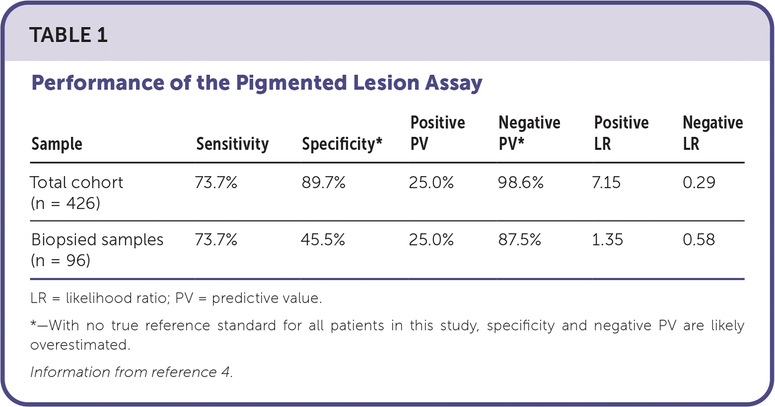
A retrospective cohort study assessed the performance of PLA testing during routine clinical use in dermatology clinics. There was no reference standard for all patients. The study included 426 pigmented lesions, 96 of which were biopsied based on clinical concern. Of the total lesions, 370 were negative on PLA testing, and 56 were positive. Of the 40 PLA-negative lesions biopsied due to high clinical concern, five (12.5%) were diagnosed as melanoma, and 16 (40%) were diagnosed as atypical melanocytic nevi. The remaining PLA-negative lesions were managed with surveillance, and none exhibited concerning clinical changes after four to six months. Of the 56 PLA-positive lesions, 14 (25%) were diagnosed as melanoma. The number needed to biopsy to detect one melanoma was 5 (19 out of 96), with a biopsy ratio of 4.1 benign lesions for each melanoma detected. For the total cohort (n = 426), PLA-negative results without a biopsy were treated as true negatives (due to low clinical concern). For the biopsied cohort, true negatives were determined by a histologic diagnosis of nonmelanoma (Table 1).4
TABLE 1. Performance of the Pigmented Lesion Assay
| Sample | Sensitivity | Specificity* | Positive PV | Negative PV* | Positive LR | Negative LR |
|---|---|---|---|---|---|---|
| Total cohort (n = 426) | 73.7% | 89.7% | 25.0% | 98.6% | 7.15 | 0.29 |
| Biopsied samples (n = 96) | 73.7% | 45.5% | 25.0% | 87.5% | 1.35 | 0.58 |
LR = likelihood ratio; PV = predictive value.
*—With no true reference standard for all patients in this study, specificity and negative PV are likely overestimated.
Information from reference 4.
Read the full article
Get immediate access, anytime, anywhere.
Choose a single article, issue, or full-access subscription.
Earn up to 14 CME credits per issue.
