

Rashes in the newborn period are common and most are benign. Infections should be suspected in newborns with pustules or vesicles, especially in those who are not well-appearing or have risk factors for congenital infection. Congenital cytomegalovirus infection can cause sensorineural hearing loss and neurodevelopmental delay. Skin manifestations of cytomegalovirus may include petechiae due to thrombocytopenia. The most common skin manifestations of early congenital syphilis are small, copper-red, maculopapular lesions located primarily on the hands and feet that peel and crust over three weeks. Erythema toxicum neonatorum and neonatal pustular melanosis are transient pustular rashes with characteristic appearance and distribution. Neonatal acne is self-limited, whereas infantile acne may benefit from treatment. Milia can be differentiated from neonatal acne by their presence at birth. Cutis marmorata and harlequin color change are transient vascular phenomena resulting from inappropriate or exaggerated dilation of capillaries and venules in response to stimuli.
Skin findings in newborns can present a diagnostic challenge in distinguishing common, benign rashes from those associated with infection, malignancy, or systemic syndromes. When clinicians evaluate the newborn rash, the most important skill is to recognize when further evaluation is necessary because early diagnosis and treatment can have a significant impact on morbidity and mortality. Part I of this article reviews the presentation, prognosis, and treatment of the most common rashes and skin changes that present during the first four weeks of life. Part II of this article, which appears in this issue of American Family Physician, discusses the identification and management of birthmarks that appear in newborns.1
SORT: KEY RECOMMENDATIONS FOR PRACTICE
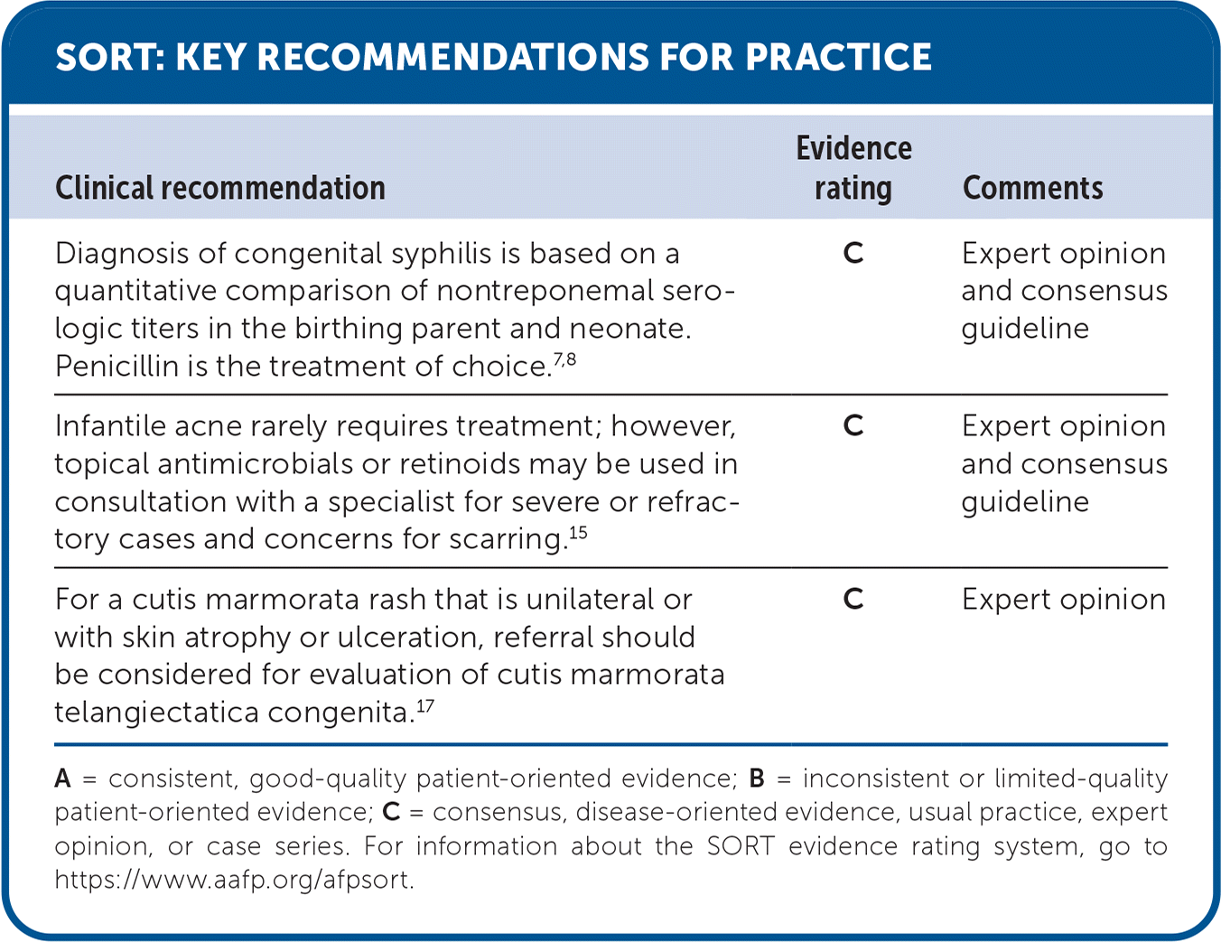
| Clinical recommendation | Evidence rating | Comments |
|---|---|---|
| Diagnosis of congenital syphilis is based on a quantitative comparison of nontreponemal serologic titers in the birthing parent and neonate. Penicillin is the treatment of choice.7,8 | C | Expert opinion and consensus guideline |
| Infantile acne rarely requires treatment; however, topical antimicrobials or retinoids may be used in consultation with a specialist for severe or refractory cases and concerns for scarring.15 | C | Expert opinion and consensus guideline |
| For a cutis marmorata rash that is unilateral or with skin atrophy or ulceration, referral should be considered for evaluation of cutis marmorata telangiectatica congenita.17 | C | Expert opinion |
A = consistent, good-quality patient-oriented evidence; B = inconsistent or limited-quality patient-oriented evidence; C = consensus, disease-oriented evidence, usual practice, expert opinion, or case series. For information about the SORT evidence rating system, go to https://www.aafp.org/afpsort.
Transient Rashes
Infectious causes of transient rashes, such as Candida infections or congenital cytomegalovirus, should be a primary consideration. A rash consistent with one of the classic benign presentations in the well-appearing newborn can be monitored for resolution. Risk factors for congenital infections should be considered before the diagnosis of a benign rash.
INFECTIOUS CAUSES
Congenital infections may present with vesicles or pustules and can generally be distinguished based on presentation (Table 1).2–8 Diffuse papular or vesicular rashes due to bacterial infections will often be associated with clinical signs of sepsis.2
Read the full article
Get immediate access, anytime, anywhere.
Choose a single article, issue, or full-access subscription.
Earn up to 14 CME credits per issue.
