

An 82-year-old man presented with pain in his left groin and difficulty walking after a fall. Radiography revealed a fracture of the left proximal femur, and surgery was scheduled. Laboratory tests showed mild anemia but no other abnormalities.
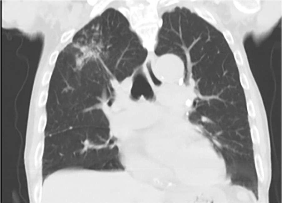
A shadow in the right upper lung field was visible on chest radiography (Figure 1). Computed tomography of the chest revealed ground-glass opacity nodules in the right upper lung field (Figure 2 and Figure 3). He did not have a history of smoking or respiratory disease.
FIGURE 1
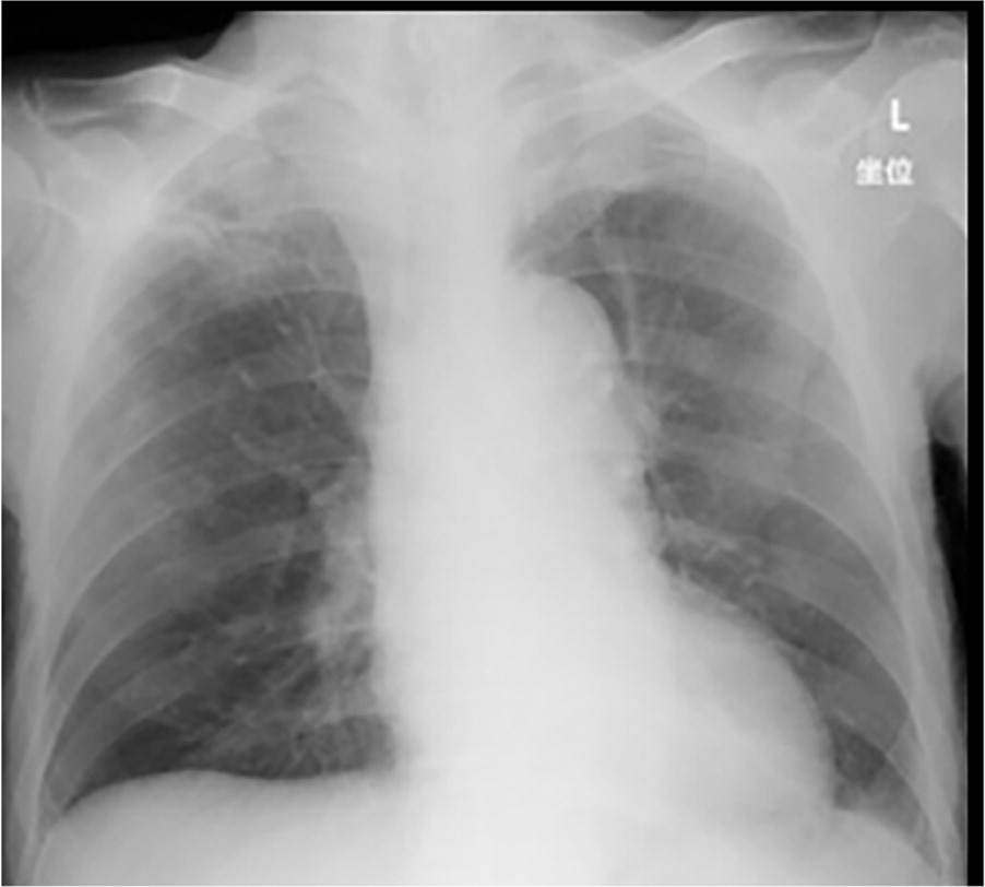
FIGURE 2
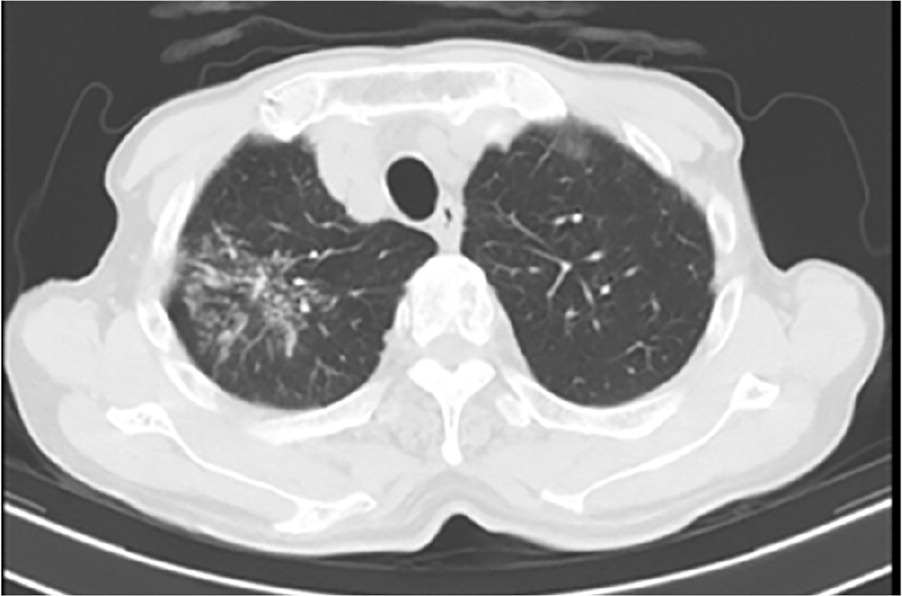
FIGURE 3
QUESTION
Based on the patient's history, physical examination, and imaging, which one of the following is the most likely diagnosis?
- A. Histoplasmosis.
- B. Lung cancer.
- C. Mucosa-associated lymphoid tissue lymphoma.
- D. Pulmonary tuberculosis.
- E. Sarcoidosis.
DISCUSSION
The answer is D: pulmonary tuberculosis. Sputum and gastric juices were examined based on the cluster of nodules in the right upper lung field. Results of polymerase chain reaction testing of the gastric juices were positive for Mycobacterium tuberculosis. Results of a T-Spot interferon-gamma release assay were also positive. The patient was diagnosed with pulmonary tuberculosis and scheduled for an antituberculosis regimen consisting of rifampicin, isoniazid, and ethambutol for 2 months and rifampicin and isoniazid for an additional 7 months.
After consultation with an orthopedic surgeon, it was determined that the patient could wait 2 weeks for surgery, and the antituberculosis regimen was started to prevent nosocomial infection. Hip replacement surgery was performed, and the patient was discharged from the hospital after 1 month of rehabilitation. While the patient was in the hospital, chest radiography and T-Spot tests were performed on his family members to ensure that the pulmonary tuberculosis had not spread.
Asymptomatic cases (latent tuberculosis) are often detected incidentally on chest radiography; despite having active tuberculosis infection, patients often have negative findings on acid-fast bacilli staining and culture.1 Radiography findings may include nodules surrounded by centrilobular or satellite micronodules in the upper lung zone, in the absence of characteristic findings such as cavitation or the tree-in-bud sign.
Histoplasmosis is a fungal infection that is endemic to the Mississippi and Ohio River Valleys. Most patients are asymptomatic. Chest radiography can reveal patchy diffuse opacities, interstitial infiltrates, and mediastinal and portal lymphadenopathy. Calcified nodules are commonly seen with previous exposure.2
Lung cancer characterized by ground-glass opacity nodules is often early carcinoma or its preinvasive lesions, atypical adenomatous hyperplasia, or adenocarcinoma in situ. Lung cancer with ground-glass opacity nodules is notable for its increasing prevalence, unique natural course, and association with lung adenocarcinoma. This disease is slow to develop and patients are usually female, nonsmokers, of Asian descent, and relatively young.3
Mucosa-associated lymphoid tissue lymphoma is the most common primary pulmonary lymphoma.4 It is often diagnosed definitively with open or thoracoscopic lung biopsy. Histologically, this group of tumors is characterized by a noninvasive lymphoplasmacytic infiltrate. Mucosa-associated lymphoid tissue lymphoma usually remains localized for prolonged periods and is responsive to locally directed therapy.
Sarcoidosis is a multisystem disorder of unknown origin and poorly understood pathogenesis that predominantly affects the lungs and intrathoracic lymph nodes.5 It is characterized by the presence of noncaseating granulomatous inflammation in involved organs. The most commonly involved organs are the lungs, lymph nodes, skin, and eyes. Bronchoscopy is most useful in diagnosing sarcoidosis except in biopsy sites such as skin or superficial lymph nodes.
Read the full article
Get immediate access, anytime, anywhere.
Choose a single article, issue, or full-access subscription.
Earn up to 14 CME credits per issue.
