

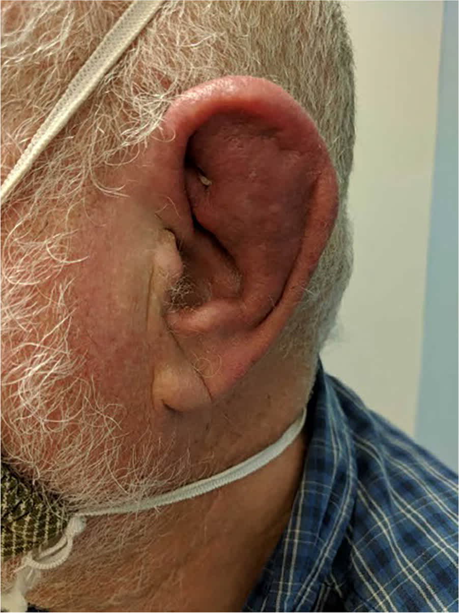
A 69-year-old man presented with a swollen left auricle. He had no improvement with a course of topical mupirocin. After 4 weeks, he reported worsening pain and swelling and could no longer tolerate sleeping on the affected side. The patient had no ear drainage, trauma, hearing changes, vertigo, or fever. His history included a similar episode 10 years earlier that resolved spontaneously. Physical examination revealed a red, tender, swollen left auricle, with sparing of the lobule (Figure 1).
FIGURE 1
QUESTION
Based on the patient's history and physical examination, which one of the following is the most likely diagnosis?
- A. Acute otitis externa.
- B. Chondrodermatitis nodularis helicis.
- C. Granulomatosis with polyangiitis.
- D. Herpes zoster oticus.
- E. Relapsing polychondritis.
DISCUSSION
The answer is E: relapsing polychondritis. This is a rare, multisystem autoimmune condition of cartilaginous structures that typically features recurrent self-limited flare-ups. Peak onset occurs between 40 and 50 years of age.1 Relapsing polychondritis is strongly associated with other concurrent autoimmune disorders. The auricle is the most common site of inflammation with sparing of the noncartilaginous lobule. Nasal chondritis and migrating nonerosive oligoarthritis are also typically present.
Relapsing polychondritis can also involve the tracheobronchial tree and cardiac connective tissues, leading to life-threatening cardiopulmonary complications. This is the leading cause of morbidity and mortality associated with the condition. Vascular, dermatologic, renal, ocular, and neurologic manifestations are also well-documented. The range of clinical manifestations at any cartilaginous site makes early diagnosis challenging. Treatment typically includes systemic steroids or another form of immunosuppression.2
Acute otitis externa is inflammation of the external ear, usually due to bacterial infection. It is not associated with lobule-sparing erythema. Topical antibiotics are typically an effective treatment.3
Chondrodermatitis nodularis helicis is a benign inflammatory condition affecting the skin and cartilage of the auricular helix, usually on the patient's habitual sleeping side. It presents as a painful, indurated nodule with subsequent ulceration.4,5
Granulomatosis with polyangiitis is a necrotizing vasculitis associated with antineutrophil cytoplasmic autoantibodies. It manifests in the ears, nose, throat, respiratory tract, and kidneys. Granulomatosis can destroy facial cartilage, although the most common symptom is recurrent sinusitis with nasal obstruction and hyposmia.6
Herpes zoster oticus (Ramsay Hunt syndrome) is a vesicular eruption of Varicella zoster virus affecting the ear. The clinical diagnosis is made when an eruption is associated with ipsilateral facial nerve palsy. Most cases present with a vesicular rash on the areas of the auricle innervated by the facial nerve.7
SUMMARY TABLE
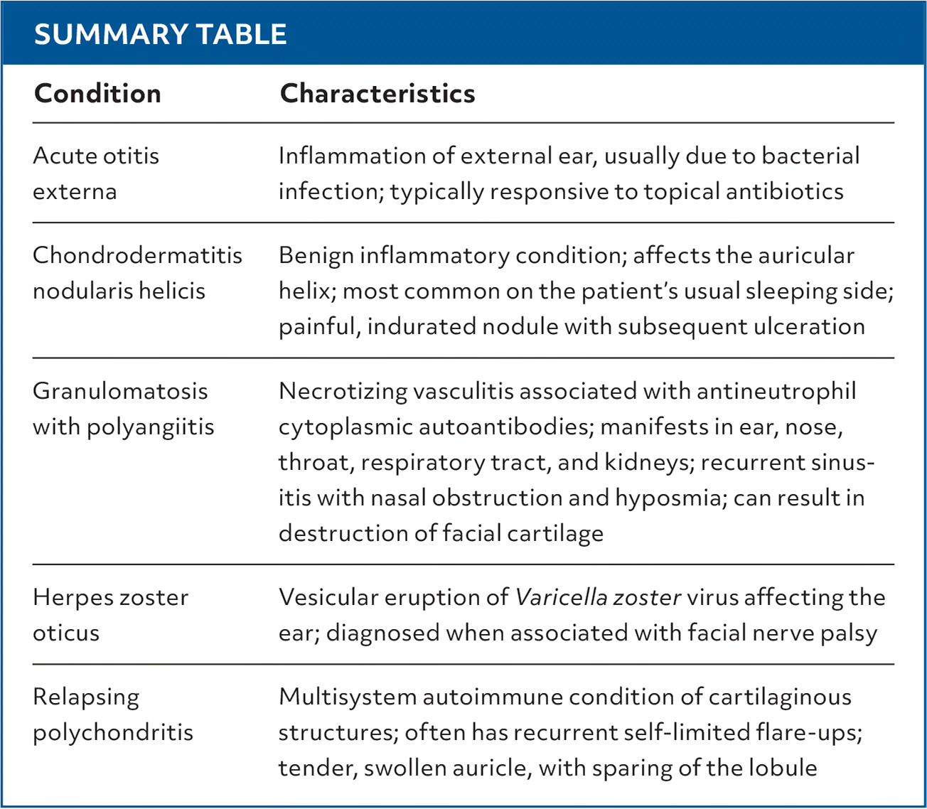
| Condition | Characteristics |
|---|---|
| Acute otitis externa | Inflammation of external ear, usually due to bacterial infection; typically responsive to topical antibiotics |
| Chondrodermatitis nodularis helicis | Benign inflammatory condition; affects the auricular helix; most common on the patient's usual sleeping side; painful, indurated nodule with subsequent ulceration |
| Granulomatosis with polyangiitis | Necrotizing vasculitis associated with antineutrophil cytoplasmic autoantibodies; manifests in ear, nose, throat, respiratory tract, and kidneys; recurrent sinustis with nasal obstruction and hyposmia; can result in destruction of facial cartilage |
| Herpes zoster oticus | Vesicular eruption of Varicella zoster virus affecting the ear; diagnosed when associated with facial nerve palsy |
| Relapsing polychondritis | Multisystem autoimmune condition of cartilaginous structures; often has recurrent self-limited flare-ups; tender, swollen auricle, with sparing of the lobule |
Read the full article
Get immediate access, anytime, anywhere.
Choose a single article, issue, or full-access subscription.
Earn up to 14 CME credits per issue.
